{"title":"Oestradiol-mediated ferroptosis defense shapes sex differences in acute kidney injury","authors":"Shengrong Wu, Li Zhuang, Boyi Gan","doi":"10.1038/s41418-025-01573-w","DOIUrl":null,"url":null,"abstract":"<p>Acute kidney injury (AKI) is a leading cause of morbidity and mortality. For decades, clinical observations and epidemiological studies have indicated that premenopausal women are less prone to AKI than men and postmenopausal women [1, 2]. However, the biological basis for this sex bias has remained largely unknown. In a recent <i>Nature</i> paper [3], Tonnus et al. provide a compelling mechanistic explanation, demonstrating that female renal tubules are inherently resistant to ferroptosis—a predominant mode of cell death involved in ischemic AKI—through oestradiol-mediated protection against this cell death.</p><p>AKI often arises from acute tubular necrosis, a pathological condition characterized by spatially propagating cell death along the nephron; this propagation has been shown to be driven, at least in part, by ferroptosis, a form of lipid peroxidation–driven cell death exacerbated by iron overload and impaired antioxidant defenses [4, 5]. Notably, previous studies reported that female mice were substantially more resistant to AKI and renal tubular ferroptosis propagation than male counterparts [6]. Using ischemia–reperfusion injury (IRI) models and ex vivo assays with isolated renal tubules, Tonnus et al. confirmed these findings and showed that ferroptotic cell death waves were prominent in male tubules but virtually absent in females; furthermore, while ferroptosis inhibitors have shown a robust protective effect in male mice, their benefit in females has been modest [3]. This resistance could not be explained by differences in the expression of classical ferroptosis regulators such as glutathione peroxidase 4 (GPX4).</p>","PeriodicalId":9731,"journal":{"name":"Cell Death and Differentiation","volume":"7 1","pages":""},"PeriodicalIF":15.4000,"publicationDate":"2025-08-28","publicationTypes":"Journal Article","fieldsOfStudy":null,"isOpenAccess":false,"openAccessPdf":"","citationCount":"0","resultStr":null,"platform":"Semanticscholar","paperid":null,"PeriodicalName":"Cell Death and Differentiation","FirstCategoryId":"99","ListUrlMain":"https://doi.org/10.1038/s41418-025-01573-w","RegionNum":1,"RegionCategory":"生物学","ArticlePicture":[],"TitleCN":null,"AbstractTextCN":null,"PMCID":null,"EPubDate":"","PubModel":"","JCR":"Q1","JCRName":"BIOCHEMISTRY & MOLECULAR BIOLOGY","Score":null,"Total":0}
引用次数: 0
Abstract
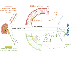
Acute kidney injury (AKI) is a leading cause of morbidity and mortality. For decades, clinical observations and epidemiological studies have indicated that premenopausal women are less prone to AKI than men and postmenopausal women [1, 2]. However, the biological basis for this sex bias has remained largely unknown. In a recent Nature paper [3], Tonnus et al. provide a compelling mechanistic explanation, demonstrating that female renal tubules are inherently resistant to ferroptosis—a predominant mode of cell death involved in ischemic AKI—through oestradiol-mediated protection against this cell death.
AKI often arises from acute tubular necrosis, a pathological condition characterized by spatially propagating cell death along the nephron; this propagation has been shown to be driven, at least in part, by ferroptosis, a form of lipid peroxidation–driven cell death exacerbated by iron overload and impaired antioxidant defenses [4, 5]. Notably, previous studies reported that female mice were substantially more resistant to AKI and renal tubular ferroptosis propagation than male counterparts [6]. Using ischemia–reperfusion injury (IRI) models and ex vivo assays with isolated renal tubules, Tonnus et al. confirmed these findings and showed that ferroptotic cell death waves were prominent in male tubules but virtually absent in females; furthermore, while ferroptosis inhibitors have shown a robust protective effect in male mice, their benefit in females has been modest [3]. This resistance could not be explained by differences in the expression of classical ferroptosis regulators such as glutathione peroxidase 4 (GPX4).
期刊介绍:
Mission, vision and values of Cell Death & Differentiation:
To devote itself to scientific excellence in the field of cell biology, molecular biology, and biochemistry of cell death and disease.
To provide a unified forum for scientists and clinical researchers
It is committed to the rapid publication of high quality original papers relating to these subjects, together with topical, usually solicited, reviews, meeting reports, editorial correspondence and occasional commentaries on controversial and scientifically informative issues.
 求助内容:
求助内容: 应助结果提醒方式:


