Natasha Patel, Truc T. Pham, Arshiya Banu, Alex Griffiths, Brett M. Paterson, George Firth, Alexander Morrell, Clíona McMahon, Nicholas J. Long, James R. Baker, Vijay Chudasama and Michelle T. Ma
{"title":"Mass spectrometric imaging and quantitative analysis of the in vivo biodistribution of trastuzumab using a rhodium(iii) sarcophagine complex","authors":"Natasha Patel, Truc T. Pham, Arshiya Banu, Alex Griffiths, Brett M. Paterson, George Firth, Alexander Morrell, Clíona McMahon, Nicholas J. Long, James R. Baker, Vijay Chudasama and Michelle T. Ma","doi":"10.1039/D5QI00731C","DOIUrl":null,"url":null,"abstract":"<p >Mass cytometry with antibodies labelled with stable metal isotopes enables both sensitive imaging and the quantification of protein expression in biological samples. Typically, these specimens are exposed to a panel of labelled antibodies <em>ex vivo</em>, after sample collection. Here, we have developed a rhodium-labelled immunoconjugate of the HER2-targeted therapeutic IgG1 antibody, trastuzumab, and evaluated its <em>in vivo</em> biodistribution using mass cytometry techniques. A Rh<small><sup>3+</sup></small> complex of a macrobicyclic sarcophagine (sar, 3,6,10,13,16,19-hexaazabicyclo[6.6.6]icosane) chelator was appended with a dibromopyridazinedione (DBPD), to produce a novel disulfide bond labelling molecule, “Rh-sar-DBPD”. Rh-sar-DBPD was site-specifically conjugated to trastuzumab <em>via</em> its four native solvent-accessible disulfide bonds, to yield a near homogeneous, well-defined and stable pyridazinedione (PD) immunoconjugate, Rh-sar-PD-trastuzumab, in which four Rh-sar-PD groups were attached per molecule of trastuzumab. Inductively coupled plasma mass spectrometry (ICP-MS) and laser ablation inductively coupled plasma mass spectrometry (LA-ICP-MS) were then applied to measure <small><sup>103</sup></small>Rh content, as a proxy for Rh-sar-PD-trastuzumab accumulation, in <em>in vitro</em> and <em>in vivo</em> studies. ICP-MS <em>in vitro</em> studies indicated HER2-mediated uptake of Rh-sar-PD-trastuzumab in HER2-expressing breast cancer cells, with LA-ICP-MS images showing intercellular heterogeneity in Rh-sar-PD-trastuzumab uptake. To study the <em>in vivo</em> biodistribution of Rh-sar-PD-trastuzumab, female NSG mice bearing orthotopic HCC1954 breast cancer tumours were administered the immunoconjugate. Quantitative ICP-MS of <small><sup>103</sup></small>Rh signal in dissected tissues indicated receptor-specific HER2-mediated uptake in tumours, as well as accumulation in the spleen and liver. Finally, LA-ICP-MS imaging analysis of tumour and ovary tissue sections showed heterogeneous uptake in HER2-expressing HCC1954 tumour cells and follicular granulosa cells of the ovaries, which are known to express growth factor receptors. To the best of our knowledge, this is the first report in which both ICP-MS and LA-ICP-MS have been used on tissue exposed to a metal-tagged antibody <em>in vivo</em>, enabling quantification of the biodistribution of the novel immunoconjugate, Rh-sar-PD-trastuzumab, in a murine model of breast cancer.</p>","PeriodicalId":79,"journal":{"name":"Inorganic Chemistry Frontiers","volume":" 19","pages":" 5688-5700"},"PeriodicalIF":6.4000,"publicationDate":"2025-08-26","publicationTypes":"Journal Article","fieldsOfStudy":null,"isOpenAccess":false,"openAccessPdf":"https://pubs.rsc.org/en/content/articlepdf/2025/qi/d5qi00731c?page=search","citationCount":"0","resultStr":null,"platform":"Semanticscholar","paperid":null,"PeriodicalName":"Inorganic Chemistry Frontiers","FirstCategoryId":"92","ListUrlMain":"https://pubs.rsc.org/en/content/articlelanding/2025/qi/d5qi00731c","RegionNum":1,"RegionCategory":"化学","ArticlePicture":[],"TitleCN":null,"AbstractTextCN":null,"PMCID":null,"EPubDate":"","PubModel":"","JCR":"Q1","JCRName":"CHEMISTRY, INORGANIC & NUCLEAR","Score":null,"Total":0}
引用次数: 0
Abstract
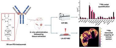
Mass cytometry with antibodies labelled with stable metal isotopes enables both sensitive imaging and the quantification of protein expression in biological samples. Typically, these specimens are exposed to a panel of labelled antibodies ex vivo, after sample collection. Here, we have developed a rhodium-labelled immunoconjugate of the HER2-targeted therapeutic IgG1 antibody, trastuzumab, and evaluated its in vivo biodistribution using mass cytometry techniques. A Rh3+ complex of a macrobicyclic sarcophagine (sar, 3,6,10,13,16,19-hexaazabicyclo[6.6.6]icosane) chelator was appended with a dibromopyridazinedione (DBPD), to produce a novel disulfide bond labelling molecule, “Rh-sar-DBPD”. Rh-sar-DBPD was site-specifically conjugated to trastuzumab via its four native solvent-accessible disulfide bonds, to yield a near homogeneous, well-defined and stable pyridazinedione (PD) immunoconjugate, Rh-sar-PD-trastuzumab, in which four Rh-sar-PD groups were attached per molecule of trastuzumab. Inductively coupled plasma mass spectrometry (ICP-MS) and laser ablation inductively coupled plasma mass spectrometry (LA-ICP-MS) were then applied to measure 103Rh content, as a proxy for Rh-sar-PD-trastuzumab accumulation, in in vitro and in vivo studies. ICP-MS in vitro studies indicated HER2-mediated uptake of Rh-sar-PD-trastuzumab in HER2-expressing breast cancer cells, with LA-ICP-MS images showing intercellular heterogeneity in Rh-sar-PD-trastuzumab uptake. To study the in vivo biodistribution of Rh-sar-PD-trastuzumab, female NSG mice bearing orthotopic HCC1954 breast cancer tumours were administered the immunoconjugate. Quantitative ICP-MS of 103Rh signal in dissected tissues indicated receptor-specific HER2-mediated uptake in tumours, as well as accumulation in the spleen and liver. Finally, LA-ICP-MS imaging analysis of tumour and ovary tissue sections showed heterogeneous uptake in HER2-expressing HCC1954 tumour cells and follicular granulosa cells of the ovaries, which are known to express growth factor receptors. To the best of our knowledge, this is the first report in which both ICP-MS and LA-ICP-MS have been used on tissue exposed to a metal-tagged antibody in vivo, enabling quantification of the biodistribution of the novel immunoconjugate, Rh-sar-PD-trastuzumab, in a murine model of breast cancer.
 求助内容:
求助内容: 应助结果提醒方式:


