Preclinical Fluorescence-Guided Imaging Leveraging Surrounding Sentinel Tumor Microenvironment Identifies High-Risk Premalignant Pancreatic Lesions

IF 10.2 1区 医学 Q1 ONCOLOGY
Shilpa Sharma, Xiaoxia Wen, Jianbo Wang, Beibei Huang, Denise Hernandez, Cong-Dat Pham, Zhiwen Liu, Susanne Je-Han. Lin, Aiko Yamaguchi, Dimitra K. Georgiou, Ryan P. Coll, H. Charles Manning
{"title":"Preclinical Fluorescence-Guided Imaging Leveraging Surrounding Sentinel Tumor Microenvironment Identifies High-Risk Premalignant Pancreatic Lesions","authors":"Shilpa Sharma, Xiaoxia Wen, Jianbo Wang, Beibei Huang, Denise Hernandez, Cong-Dat Pham, Zhiwen Liu, Susanne Je-Han. Lin, Aiko Yamaguchi, Dimitra K. Georgiou, Ryan P. Coll, H. Charles Manning","doi":"10.1158/1078-0432.ccr-25-1092","DOIUrl":null,"url":null,"abstract":"Purpose: With surgery being the only potential cure for pancreatic cancer, high-risk premalignant pancreatic lesions often go unnoticed by palpation or white light visualization, leading to recurrence. We asked whether near-infrared fluorescence imaging of tumor-associated inflammation could identify high-risk premalignant lesions, leveraging the tumor microenvironment (TME) as a sentinel of local disease and, thus, enhance surgery outcomes. Experimental Design: Fluorescence-guided surgery was performed on genetically engineered mice (Ptf1a-Cre; LSL-KrasG12D/+; Smad4flox/flox [KSC]) at discrete stages of disease progression, histologically confirmed high-risk, premalignant lesions in postnatal mice to locally advanced pancreatic tumors in adults, using the imaging agent V-1520, a translocator protein (TSPO) ligand. Age-matched wild-type littermates were used as controls, while Ptf1a-Cre; LSL-KrasG12D/+(KC) mice modeled pancreatitis and precursors of low penetrance. Localization of V-1520 and tumor-associated macrophages amongst the TME was detected by immunofluorescence imaging. Results: V-1520 exhibited robust accumulation in the pancreata of KSC mice from the early postnatal stage. Increased accumulation was observed in the pancreata of adolescent- and adult-aged mice with greater ductal lesion and stromal burden. Confocal microscopy of ex vivo pancreas specimens co-localized V-1520 accumulation primarily with CD68-expressing macrophage in KSC mice. Unlike the pancreata of KSC mice, accumulation of V-1520 did not exceed background levels in the pancreata of KC mice with pancreatitis. Conclusion: V-1520 exhibited differential accumulation in pancreatic cancer-associated inflammation compared to pancreatitis. Given the robust tracer uptake in tissues associated with early yet high-risk lesions, we envision V-1520 could enhance surgical resection and reduce the potential for recurrence from residual disease.","PeriodicalId":10279,"journal":{"name":"Clinical Cancer Research","volume":"22 1","pages":""},"PeriodicalIF":10.2000,"publicationDate":"2025-08-25","publicationTypes":"Journal Article","fieldsOfStudy":null,"isOpenAccess":false,"openAccessPdf":"","citationCount":"0","resultStr":null,"platform":"Semanticscholar","paperid":null,"PeriodicalName":"Clinical Cancer Research","FirstCategoryId":"3","ListUrlMain":"https://doi.org/10.1158/1078-0432.ccr-25-1092","RegionNum":1,"RegionCategory":"医学","ArticlePicture":[],"TitleCN":null,"AbstractTextCN":null,"PMCID":null,"EPubDate":"","PubModel":"","JCR":"Q1","JCRName":"ONCOLOGY","Score":null,"Total":0}
引用次数: 0

Abstract

Purpose: With surgery being the only potential cure for pancreatic cancer, high-risk premalignant pancreatic lesions often go unnoticed by palpation or white light visualization, leading to recurrence. We asked whether near-infrared fluorescence imaging of tumor-associated inflammation could identify high-risk premalignant lesions, leveraging the tumor microenvironment (TME) as a sentinel of local disease and, thus, enhance surgery outcomes. Experimental Design: Fluorescence-guided surgery was performed on genetically engineered mice (Ptf1a-Cre; LSL-KrasG12D/+; Smad4flox/flox [KSC]) at discrete stages of disease progression, histologically confirmed high-risk, premalignant lesions in postnatal mice to locally advanced pancreatic tumors in adults, using the imaging agent V-1520, a translocator protein (TSPO) ligand. Age-matched wild-type littermates were used as controls, while Ptf1a-Cre; LSL-KrasG12D/+(KC) mice modeled pancreatitis and precursors of low penetrance. Localization of V-1520 and tumor-associated macrophages amongst the TME was detected by immunofluorescence imaging. Results: V-1520 exhibited robust accumulation in the pancreata of KSC mice from the early postnatal stage. Increased accumulation was observed in the pancreata of adolescent- and adult-aged mice with greater ductal lesion and stromal burden. Confocal microscopy of ex vivo pancreas specimens co-localized V-1520 accumulation primarily with CD68-expressing macrophage in KSC mice. Unlike the pancreata of KSC mice, accumulation of V-1520 did not exceed background levels in the pancreata of KC mice with pancreatitis. Conclusion: V-1520 exhibited differential accumulation in pancreatic cancer-associated inflammation compared to pancreatitis. Given the robust tracer uptake in tissues associated with early yet high-risk lesions, we envision V-1520 could enhance surgical resection and reduce the potential for recurrence from residual disease.
利用周围前哨肿瘤微环境的临床前荧光引导成像识别高危癌前胰腺病变
目的:手术是胰腺癌唯一可能的治疗方法,但胰腺高危癌前病变常被触诊或白光显像所忽视,从而导致复发。我们询问肿瘤相关炎症的近红外荧光成像是否可以识别高风险的癌前病变,利用肿瘤微环境(TME)作为局部疾病的哨兵,从而提高手术效果。实验设计:使用显像剂V-1520(一种转运蛋白(TSPO)配体),在疾病进展的不同阶段对基因工程小鼠(Ptf1a-Cre、LSL-KrasG12D/+、Smad4flox/flox [KSC])进行荧光引导手术,组织学证实的高风险、产后小鼠的癌前病变到成年小鼠的局部晚期胰腺肿瘤。以年龄匹配的野生型幼崽为对照,Ptf1a-Cre;LSL-KrasG12D/+(KC)小鼠模型胰腺炎和低外显率前体。免疫荧光成像检测V-1520和肿瘤相关巨噬细胞在TME中的定位。结果:V-1520从出生后早期开始在KSC小鼠胰腺中表现出强劲的积累。在导管病变和间质负担较大的青春期和成年小鼠胰腺中观察到积累增加。离体胰腺标本的共聚焦显微镜在KSC小鼠中主要与表达cd68的巨噬细胞共定位V-1520积累。与KSC小鼠的胰腺不同,患有胰腺炎的KC小鼠的胰腺中V-1520的积累没有超过背景水平。结论:与胰腺炎相比,V-1520在胰腺癌相关炎症中表现出不同的积累。鉴于示踪剂在与早期高风险病变相关的组织中的强劲摄取,我们设想V-1520可以增强手术切除并减少残留疾病复发的可能性。
本文章由计算机程序翻译,如有差异,请以英文原文为准。
求助全文
约1分钟内获得全文 求助全文
来源期刊
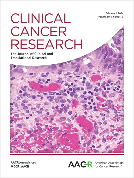
Clinical Cancer Research
Clinical Cancer Research 医学-肿瘤学
CiteScore
20.10
自引率
1.70%
发文量
1207
审稿时长
2.1 months
期刊介绍: Clinical Cancer Research is a journal focusing on groundbreaking research in cancer, specifically in the areas where the laboratory and the clinic intersect. Our primary interest lies in clinical trials that investigate novel treatments, accompanied by research on pharmacology, molecular alterations, and biomarkers that can predict response or resistance to these treatments. Furthermore, we prioritize laboratory and animal studies that explore new drugs and targeted agents with the potential to advance to clinical trials. We also encourage research on targetable mechanisms of cancer development, progression, and metastasis.
×
引用
GB/T 7714-2015
复制
MLA
复制
APA
复制
导出至
BibTeX EndNote RefMan NoteFirst NoteExpress
×
提示
您的信息不完整,为了账户安全,请先补充。
现在去补充
×
提示
您因"违规操作"
具体请查看互助需知
我知道了
×
提示
确定
请完成安全验证×
copy
已复制链接
快去分享给好友吧!
我知道了
右上角分享
点击右上角分享
0
联系我们:info@booksci.cn Book学术提供免费学术资源搜索服务,方便国内外学者检索中英文文献。致力于提供最便捷和优质的服务体验。 Copyright © 2023 布克学术 All rights reserved.
京ICP备2023020795号-1
ghs 京公网安备 11010802042870号
Book学术文献互助
Book学术文献互助群
群 号:604180095
Book学术官方微信