Development and Validation of a Computed Tomography-based Radiomics Nomogram for Diagnosing Cervical Lymph Node Metastasis in Oropharyngeal Squamous Cell Carcinomas.
Ran Zhao, Changdong Ma, Jingmin Zou, Xueli Fang, Qiang Wu, Chao Kong, Changsheng Ma, Kai Liu
{"title":"Development and Validation of a Computed Tomography-based Radiomics Nomogram for Diagnosing Cervical Lymph Node Metastasis in Oropharyngeal Squamous Cell Carcinomas.","authors":"Ran Zhao, Changdong Ma, Jingmin Zou, Xueli Fang, Qiang Wu, Chao Kong, Changsheng Ma, Kai Liu","doi":"10.1016/j.adro.2025.101844","DOIUrl":null,"url":null,"abstract":"<p><strong>Purpose: </strong>To construct and validate a radiomic nomogram based on computed tomography (CT) scan data to diagnose lymph node (LN) metastasis (LNM) in patients with oropharyngeal squamous cell carcinoma (OPSCC) and compare it with a model based on CT scan signs recognized by the naked eye.</p><p><strong>Methods and materials: </strong>Data from patients who visited the authors' hospital between January 2018 and February 2023 were retrospectively reviewed. Eighty-six patients with OPSCC contributed 116 LNs, which were randomly divided into training and test sets. Radiologists derived CT signs characteristic of each LN by visually reviewing CT scan images. The radiomics features of LNs were extracted using \"3Dslicer\" (https://www.slicer.org), and the least absolute shrinkage and selection operator method was used to reduce the dimensions and establish radiomics tags. A CT scan-based radiomic nomogram was constructed and validated. The performance levels of the radiomics nomogram, radiomics signature, and CT-sign model were evaluated according to the area under the receiver operating characteristic curve (AUC) values.</p><p><strong>Results: </strong>CT signs (central necrosis, extensive necrosis, and LN accumulation) exhibited significant differences between the LN-negative and LN-positive groups. For each CT scan, 851 3-dimensional features were extracted from the cervical LN region. Eight of the most pertinent radiomic features were selected using dimensionality reduction to create radiomic tags. The radiomics nomogram incorporating the CT signs and radiomics signature demonstrated favorable predictive value for diagnosing LNM in patients with OPSCC, with the area under the receiver operating characteristic curve values of 0.983 and 0.919 for the training and test sets, respectively.</p><p><strong>Conclusion: </strong>The CT scan-based radiomics nomogram demonstrated good diagnostic utility for LNM in OPSCC and may optimize clinical decision-making. To validate our findings, future studies should consider conducting larger-scale experiments and include external validation sets to confirm the broader applicability of our results.</p>","PeriodicalId":7390,"journal":{"name":"Advances in Radiation Oncology","volume":"10 9","pages":"101844"},"PeriodicalIF":2.7000,"publicationDate":"2025-07-01","publicationTypes":"Journal Article","fieldsOfStudy":null,"isOpenAccess":false,"openAccessPdf":"https://www.ncbi.nlm.nih.gov/pmc/articles/PMC12351180/pdf/","citationCount":"0","resultStr":null,"platform":"Semanticscholar","paperid":null,"PeriodicalName":"Advances in Radiation Oncology","FirstCategoryId":"1085","ListUrlMain":"https://doi.org/10.1016/j.adro.2025.101844","RegionNum":0,"RegionCategory":null,"ArticlePicture":[],"TitleCN":null,"AbstractTextCN":null,"PMCID":null,"EPubDate":"2025/9/1 0:00:00","PubModel":"eCollection","JCR":"Q3","JCRName":"ONCOLOGY","Score":null,"Total":0}
引用次数: 0
Abstract
Purpose: To construct and validate a radiomic nomogram based on computed tomography (CT) scan data to diagnose lymph node (LN) metastasis (LNM) in patients with oropharyngeal squamous cell carcinoma (OPSCC) and compare it with a model based on CT scan signs recognized by the naked eye.
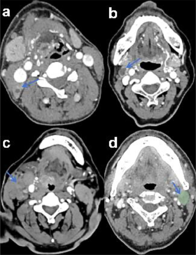
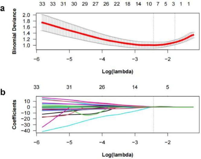
Methods and materials: Data from patients who visited the authors' hospital between January 2018 and February 2023 were retrospectively reviewed. Eighty-six patients with OPSCC contributed 116 LNs, which were randomly divided into training and test sets. Radiologists derived CT signs characteristic of each LN by visually reviewing CT scan images. The radiomics features of LNs were extracted using "3Dslicer" (https://www.slicer.org), and the least absolute shrinkage and selection operator method was used to reduce the dimensions and establish radiomics tags. A CT scan-based radiomic nomogram was constructed and validated. The performance levels of the radiomics nomogram, radiomics signature, and CT-sign model were evaluated according to the area under the receiver operating characteristic curve (AUC) values.
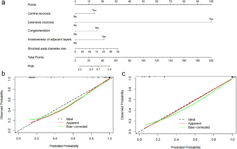
Results: CT signs (central necrosis, extensive necrosis, and LN accumulation) exhibited significant differences between the LN-negative and LN-positive groups. For each CT scan, 851 3-dimensional features were extracted from the cervical LN region. Eight of the most pertinent radiomic features were selected using dimensionality reduction to create radiomic tags. The radiomics nomogram incorporating the CT signs and radiomics signature demonstrated favorable predictive value for diagnosing LNM in patients with OPSCC, with the area under the receiver operating characteristic curve values of 0.983 and 0.919 for the training and test sets, respectively.
Conclusion: The CT scan-based radiomics nomogram demonstrated good diagnostic utility for LNM in OPSCC and may optimize clinical decision-making. To validate our findings, future studies should consider conducting larger-scale experiments and include external validation sets to confirm the broader applicability of our results.
期刊介绍:
The purpose of Advances is to provide information for clinicians who use radiation therapy by publishing: Clinical trial reports and reanalyses. Basic science original reports. Manuscripts examining health services research, comparative and cost effectiveness research, and systematic reviews. Case reports documenting unusual problems and solutions. High quality multi and single institutional series, as well as other novel retrospective hypothesis generating series. Timely critical reviews on important topics in radiation oncology, such as side effects. Articles reporting the natural history of disease and patterns of failure, particularly as they relate to treatment volume delineation. Articles on safety and quality in radiation therapy. Essays on clinical experience. Articles on practice transformation in radiation oncology, in particular: Aspects of health policy that may impact the future practice of radiation oncology. How information technology, such as data analytics and systems innovations, will change radiation oncology practice. Articles on imaging as they relate to radiation therapy treatment.
 求助内容:
求助内容: 应助结果提醒方式:


