Hella Sand, Jens Edmund, Ane Appelt, Patrick Wohlfahrt, Vicki Trier Taasti, Laurids Østergaard Poulsen, Jimmi Søndergaard, Martin Skovmos Nielsen
{"title":"Combined single- and dual-energy CT workflow for dose calculation in radiotherapy.","authors":"Hella Sand, Jens Edmund, Ane Appelt, Patrick Wohlfahrt, Vicki Trier Taasti, Laurids Østergaard Poulsen, Jimmi Søndergaard, Martin Skovmos Nielsen","doi":"10.2340/1651-226X.2025.43827","DOIUrl":null,"url":null,"abstract":"<p><strong>Background and purpose: </strong>Dual-energy computed tomography (DECT) is increasingly used in radiotherapy delineation due to its enhanced soft tissue contrast. DECT also supports direct dose calculation. However, as most current DECT scanners allow for use in only certain body regions, conventional single-energy computed tomography (SECT) is still needed for some patients. A safe clinical introduction of DECT thus requires a combined workflow. This study therefore investigates whether a unified Hounsfield look-up table (HLUT) can be applied across SECT and DECT reconstructions. Patient/material and methods: A Gammex Advanced Electron Density phantom containing tissue-equivalent inserts was scanned using SECT (70-140 kVp and Sn100-Sn140 kVp, Sn meaning tin-filtered) and dual-spiral DECT to identify matching HLUTs for three SECT methods, including a standard reconstruction (only 120 kVp; Method 1), and kVp-independent reconstructions providing mass density (MD; Method 2) or relative electron density (RED; Method 3). Dose agreement was subsequently tested on two anthropomorphic phantoms. For each SECT method, DECT reconstructions were compared through voxel-wise analysis of computed tomography (CT) numbers, and by performing dose calculations in three anatomical regions: head, thorax, and abdomen/pelvis.</p><p><strong>Results: </strong>Across all three SECT methods, DECT reconstructions with acceptable clinical CT number agreement were identified. Corresponding dose calculations between SECT- and DECT-based plans showed minimal differences.</p><p><strong>Interpretation: </strong>This phantom study demonstrates that a unified HLUT can be applied across SECT and DECT using standard 120 kVp, MD, or RED reconstructions. This approach may streamline clinical workflows and support a safe and practical transition to DECT-based treatment planning.</p>","PeriodicalId":7110,"journal":{"name":"Acta Oncologica","volume":"64 ","pages":"1079-1086"},"PeriodicalIF":2.7000,"publicationDate":"2025-08-18","publicationTypes":"Journal Article","fieldsOfStudy":null,"isOpenAccess":false,"openAccessPdf":"https://www.ncbi.nlm.nih.gov/pmc/articles/PMC12371750/pdf/","citationCount":"0","resultStr":null,"platform":"Semanticscholar","paperid":null,"PeriodicalName":"Acta Oncologica","FirstCategoryId":"3","ListUrlMain":"https://doi.org/10.2340/1651-226X.2025.43827","RegionNum":3,"RegionCategory":"医学","ArticlePicture":[],"TitleCN":null,"AbstractTextCN":null,"PMCID":null,"EPubDate":"","PubModel":"","JCR":"Q3","JCRName":"ONCOLOGY","Score":null,"Total":0}
引用次数: 0
Abstract
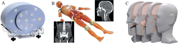
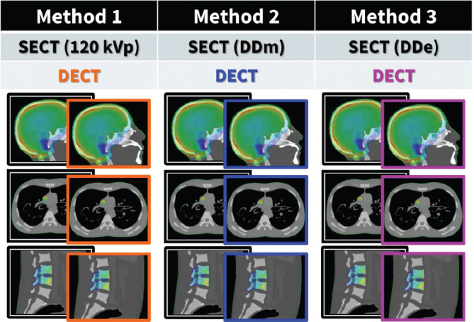
Background and purpose: Dual-energy computed tomography (DECT) is increasingly used in radiotherapy delineation due to its enhanced soft tissue contrast. DECT also supports direct dose calculation. However, as most current DECT scanners allow for use in only certain body regions, conventional single-energy computed tomography (SECT) is still needed for some patients. A safe clinical introduction of DECT thus requires a combined workflow. This study therefore investigates whether a unified Hounsfield look-up table (HLUT) can be applied across SECT and DECT reconstructions. Patient/material and methods: A Gammex Advanced Electron Density phantom containing tissue-equivalent inserts was scanned using SECT (70-140 kVp and Sn100-Sn140 kVp, Sn meaning tin-filtered) and dual-spiral DECT to identify matching HLUTs for three SECT methods, including a standard reconstruction (only 120 kVp; Method 1), and kVp-independent reconstructions providing mass density (MD; Method 2) or relative electron density (RED; Method 3). Dose agreement was subsequently tested on two anthropomorphic phantoms. For each SECT method, DECT reconstructions were compared through voxel-wise analysis of computed tomography (CT) numbers, and by performing dose calculations in three anatomical regions: head, thorax, and abdomen/pelvis.
Results: Across all three SECT methods, DECT reconstructions with acceptable clinical CT number agreement were identified. Corresponding dose calculations between SECT- and DECT-based plans showed minimal differences.
Interpretation: This phantom study demonstrates that a unified HLUT can be applied across SECT and DECT using standard 120 kVp, MD, or RED reconstructions. This approach may streamline clinical workflows and support a safe and practical transition to DECT-based treatment planning.
期刊介绍:
Acta Oncologica is a journal for the clinical oncologist and accepts articles within all fields of clinical cancer research. Articles on tumour pathology, experimental oncology, radiobiology, cancer epidemiology and medical radio physics are also welcome, especially if they have a clinical aim or interest. Scientific articles on cancer nursing and psychological or social aspects of cancer are also welcomed. Extensive material may be published as Supplements, for which special conditions apply.


 求助内容:
求助内容: 应助结果提醒方式:


