Fourier-based multiple-slice reconstruction in cryo-electron tomography
IF 2
3区 工程技术
Q2 MICROSCOPY
引用次数: 0
Abstract
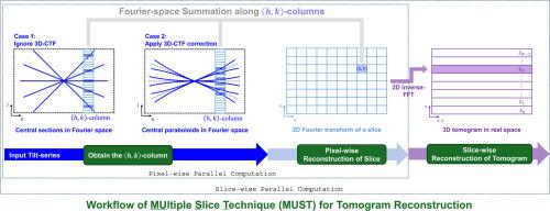
A tomogram is reconstructed from the micrographs of the tilt series using cryo-electron tomography (cryoET). Reconstruction frequently integrates image processing steps, such as filtering and contrast transfer function (CTF) correction, to support the downstream analysis of cellular and viral structures. Most image processing steps are based on Fourier space analysis, which is theoretically more efficient to be implemented in Fourier space than in real space. However, the substantial dimensions of tomograms present significant challenges for reconstruction and processing in Fourier space. Consequently, real-space reconstruction is prevalent in current practice. In this study, we proposed a Fourier-space algorithm for tomogram reconstruction, named MUltiple Slice Technique (MUST). MUST considers a tomogram composed of multiple parallel slices, with each slice independently reconstructed in Fourier space. A weighting strategy was used to enable MUST to achieve reconstruction compatible with real-space methods, including weighted back-projection (WBP) and the simultaneous iterative reconstruction technique (SIRT). A three-dimensional CTF model was formulated as pairs of conjugate central paraboloids in Fourier space and subsequently implemented for CTF correction in MUST. Alias-free reconstruction and pixel-level parallel computation are key features of MUST, demonstrated through tomogram-based subtomogram averaging at near-atomic resolutions.
低温电子断层扫描中基于傅里叶的多层重建
利用低温电子断层扫描(cryoET)从倾斜序列的显微照片重建层析图。重建通常集成图像处理步骤,如滤波和对比度传递函数(CTF)校正,以支持细胞和病毒结构的下游分析。大多数图像处理步骤都是基于傅里叶空间分析,理论上在傅里叶空间中实现比在实际空间中实现更有效。然而,层析图的大尺寸对傅里叶空间的重建和处理提出了重大挑战。因此,在当前的实践中,实空间重构是非常普遍的。在本研究中,我们提出了一种用于层析图重建的傅里叶空间算法,称为多重切片技术(MUltiple Slice Technique, MUST)。MUST考虑由多个平行切片组成的层析图,每个切片在傅里叶空间中独立重建。采用加权策略使MUST能够实现与实空间方法(包括加权反投影(WBP)和同步迭代重建技术(SIRT))兼容的重建。三维CTF模型在傅里叶空间中被表述为一对共轭中心抛物面,随后在MUST中实现CTF校正。无别名重建和像素级并行计算是MUST的关键特征,通过近原子分辨率的基于层析图的子层析图平均来证明。
本文章由计算机程序翻译,如有差异,请以英文原文为准。
求助全文
约1分钟内获得全文
求助全文
来源期刊

Ultramicroscopy
工程技术-显微镜技术
CiteScore
4.60
自引率
13.60%
发文量
117
审稿时长
5.3 months
期刊介绍:
Ultramicroscopy is an established journal that provides a forum for the publication of original research papers, invited reviews and rapid communications. The scope of Ultramicroscopy is to describe advances in instrumentation, methods and theory related to all modes of microscopical imaging, diffraction and spectroscopy in the life and physical sciences.
 求助内容:
求助内容: 应助结果提醒方式:


