Towards cardiac MRI foundation models: Comprehensive visual-tabular representations for whole-heart assessment and beyond

IF 11.8 1区 医学 Q1 COMPUTER SCIENCE, ARTIFICIAL INTELLIGENCE
Yundi Zhang , Paul Hager , Che Liu , Suprosanna Shit , Chen Chen , Daniel Rueckert , Jiazhen Pan
{"title":"Towards cardiac MRI foundation models: Comprehensive visual-tabular representations for whole-heart assessment and beyond","authors":"Yundi Zhang ,&nbsp;Paul Hager ,&nbsp;Che Liu ,&nbsp;Suprosanna Shit ,&nbsp;Chen Chen ,&nbsp;Daniel Rueckert ,&nbsp;Jiazhen Pan","doi":"10.1016/j.media.2025.103756","DOIUrl":null,"url":null,"abstract":"<div><div>Cardiac magnetic resonance (CMR) imaging is the gold standard for non-invasive cardiac assessment, offering rich spatio-temporal views of the heart’s anatomy and physiology. Patient-level health factors, such as demographics, metabolic, and lifestyle, are known to substantially influence cardiovascular health and disease risk, yet remain uncaptured by CMR alone. To holistically understand cardiac health and to enable the best possible interpretation of an individual’s disease risk, CMR and patient-level factors must be jointly exploited within an integrated framework. Recent multi-modal approaches have begun to bridge this gap, yet they often rely on limited spatio-temporal data and focus on isolated clinical tasks, thereby hindering the development of a comprehensive representation for cardiac/health evaluation.</div><div>To overcome these limitations, we introduce <em>ViTa</em>, a step toward foundation models that delivers a comprehensive representation of the heart and a precise interpretation of individual disease risk. Leveraging data from 42,000 UK Biobank participants, ViTa integrates 3D+T cine stacks from short-axis and long-axis views, enabling a complete capture of the cardiac cycle. These imaging data are then fused with detailed tabular patient-level factors, enabling context-aware insights. This multi-modal paradigm supports a wide spectrum of downstream tasks, including cardiac phenotype and physiological feature prediction, segmentation, and classification of cardiac/metabolic diseases within a single unified framework. By learning a shared latent representation that bridges rich imaging features and patient context, ViTa moves beyond traditional, task-specific models toward a universal, patient-specific understanding of cardiac health, highlighting its potential to advance clinical utility and scalability in cardiac analysis. <span><span><sup>2</sup></span></span></div></div>","PeriodicalId":18328,"journal":{"name":"Medical image analysis","volume":"107 ","pages":"Article 103756"},"PeriodicalIF":11.8000,"publicationDate":"2025-08-11","publicationTypes":"Journal Article","fieldsOfStudy":null,"isOpenAccess":false,"openAccessPdf":"","citationCount":"0","resultStr":null,"platform":"Semanticscholar","paperid":null,"PeriodicalName":"Medical image analysis","FirstCategoryId":"5","ListUrlMain":"https://www.sciencedirect.com/science/article/pii/S1361841525003032","RegionNum":1,"RegionCategory":"医学","ArticlePicture":[],"TitleCN":null,"AbstractTextCN":null,"PMCID":null,"EPubDate":"","PubModel":"","JCR":"Q1","JCRName":"COMPUTER SCIENCE, ARTIFICIAL INTELLIGENCE","Score":null,"Total":0}
引用次数: 0

Abstract

Cardiac magnetic resonance (CMR) imaging is the gold standard for non-invasive cardiac assessment, offering rich spatio-temporal views of the heart’s anatomy and physiology. Patient-level health factors, such as demographics, metabolic, and lifestyle, are known to substantially influence cardiovascular health and disease risk, yet remain uncaptured by CMR alone. To holistically understand cardiac health and to enable the best possible interpretation of an individual’s disease risk, CMR and patient-level factors must be jointly exploited within an integrated framework. Recent multi-modal approaches have begun to bridge this gap, yet they often rely on limited spatio-temporal data and focus on isolated clinical tasks, thereby hindering the development of a comprehensive representation for cardiac/health evaluation.
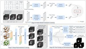
To overcome these limitations, we introduce ViTa, a step toward foundation models that delivers a comprehensive representation of the heart and a precise interpretation of individual disease risk. Leveraging data from 42,000 UK Biobank participants, ViTa integrates 3D+T cine stacks from short-axis and long-axis views, enabling a complete capture of the cardiac cycle. These imaging data are then fused with detailed tabular patient-level factors, enabling context-aware insights. This multi-modal paradigm supports a wide spectrum of downstream tasks, including cardiac phenotype and physiological feature prediction, segmentation, and classification of cardiac/metabolic diseases within a single unified framework. By learning a shared latent representation that bridges rich imaging features and patient context, ViTa moves beyond traditional, task-specific models toward a universal, patient-specific understanding of cardiac health, highlighting its potential to advance clinical utility and scalability in cardiac analysis. 2

Abstract Image

Abstract Image

心脏MRI基础模型:全心评估及其他方面的综合可视化表表示
心脏磁共振(CMR)成像是无创心脏评估的金标准,提供了丰富的心脏解剖和生理的时空视图。患者层面的健康因素,如人口统计学、代谢和生活方式,已知对心血管健康和疾病风险有重大影响,但CMR仍未单独捕获。为了全面了解心脏健康,并尽可能对个体疾病风险做出最好的解释,CMR和患者层面的因素必须在一个综合框架内共同利用。最近的多模式方法已经开始弥补这一差距,但它们往往依赖于有限的时空数据,并侧重于孤立的临床任务,从而阻碍了心脏/健康评估综合表现的发展。为了克服这些限制,我们推出了ViTa,这是向基础模型迈出的一步,它提供了心脏的全面代表和对个体疾病风险的精确解释。利用来自42000名英国生物银行参与者的数据,ViTa集成了来自短轴和长轴视图的3D+T视频堆栈,能够完整捕获心脏周期。然后将这些成像数据与详细的表格患者级因素融合在一起,从而实现上下文感知的见解。这种多模式模式支持广泛的下游任务,包括心脏表型和生理特征预测、心脏/代谢疾病的分割和分类在一个统一的框架内。通过学习连接丰富的成像特征和患者背景的共享潜在表示,ViTa超越了传统的、特定任务的模型,走向了对心脏健康的普遍、特定患者的理解,突出了其在心脏分析中推进临床应用和可扩展性的潜力。2
本文章由计算机程序翻译,如有差异,请以英文原文为准。
求助全文
约1分钟内获得全文 求助全文
来源期刊
Medical image analysis
Medical image analysis 工程技术-工程:生物医学
CiteScore
22.10
自引率
6.40%
发文量
309
审稿时长
6.6 months
期刊介绍: Medical Image Analysis serves as a platform for sharing new research findings in the realm of medical and biological image analysis, with a focus on applications of computer vision, virtual reality, and robotics to biomedical imaging challenges. The journal prioritizes the publication of high-quality, original papers contributing to the fundamental science of processing, analyzing, and utilizing medical and biological images. It welcomes approaches utilizing biomedical image datasets across all spatial scales, from molecular/cellular imaging to tissue/organ imaging.
×
引用
GB/T 7714-2015
复制
MLA
复制
APA
复制
导出至
BibTeX EndNote RefMan NoteFirst NoteExpress
×
提示
您的信息不完整,为了账户安全,请先补充。
现在去补充
×
提示
您因"违规操作"
具体请查看互助需知
我知道了
×
提示
确定
请完成安全验证×
copy
已复制链接
快去分享给好友吧!
我知道了
右上角分享
点击右上角分享
0
联系我们:info@booksci.cn Book学术提供免费学术资源搜索服务,方便国内外学者检索中英文文献。致力于提供最便捷和优质的服务体验。 Copyright © 2023 布克学术 All rights reserved.
京ICP备2023020795号-1
ghs 京公网安备 11010802042870号
Book学术文献互助
Book学术文献互助群
群 号:604180095
Book学术官方微信