{"title":"Association Between Lateral Femoral Condyle Height and Depth Ratio, Medial Tibial Plateau Slope, and Isolated Posterior Cruciate Ligament Rupture.","authors":"Kaijie Qiu, Canlong Wang, Linxiang Cheng, Yuxuan Zou, Chenchen Zhao, Zhaopeng Tang, Yang Fei, Jia Geng, Weiliang Shen, Guang Yang, Zongyou Pan","doi":"10.1177/23259671251356626","DOIUrl":null,"url":null,"abstract":"<p><strong>Background: </strong>The morphological features of the femoral condyles have recently attracted attention as a potential risk factor for knee injuries. However, little is known about whether the femoral condylar morphology is related to posterior cruciate ligament (PCL) injury.</p><p><strong>Purpose: </strong>To investigate whether the morphological characteristics of the femoral condyle are risk factors for isolated PCL rupture.</p><p><strong>Study design: </strong>Case-control study; Level of evidence, 3.</p><p><strong>Methods: </strong>From the patients who visited the outpatient clinic of our hospital between 2012 and 2022, we included 78 patients with isolated PCL ruptures and 78 age- and sex-matched controls with knee injury but no structural damage evident on magnetic resonance imaging (MRI). The following parameters were assessed using MRI: the lateral femoral condyle and medial femoral condyle height and depth ratio (LFC-H/D, MFC-H/D), notch width index, intercondylar notch angle, the lateral tibial plateau slope, and medial tibial plateau slope (MTPS), and medial tibial depth. Values were compared between these 2 groups using the independent <i>t</i> test and the Mann-Whitney <i>U</i> test. Univariate logistic regression analysis was subsequently performed to identify independent risk factors. Receiver operating characteristic curves were generated for the morphological indicators and the combination of risk factors.</p><p><strong>Results: </strong>Patients in the isolated PCL rupture group had significantly lower LFC-H/D (0.49 vs 0.52; <i>P</i> < .001) and MTPS (7.34 vs 8.81; <i>P</i> = .012) values compared with the control group. In sex-specific analyses, both male and female patients with isolated PCL rupture had a significantly lower LFC-H/D (female patients: 0.46 vs 0.52; <i>P</i> < .001; male patients: 0.51 vs 0.52; <i>P</i> = .035). In addition, male patients with PCL rupture had a smaller MTPS (6.74 mm vs 8.79 mm; <i>P</i> = .004). Univariate logistic regression analysis further validated LFC-H/D (odds ratio [OR], <0.001; <i>P</i> < .001) and MTPS (OR, 0.889; <i>P</i> = .014) as risk factors for isolated PCL rupture.</p><p><strong>Conclusion: </strong>A decreased LFC-H/D and a reduced MTPS were identified as risk factors for isolated PCL rupture. Sex-specific analysis further suggested that a decreased LFC-H/D was a risk factor for isolated PCL ruptures in both male and female patients, whereas a lower MTPS was a risk factor exclusively in male patients.</p>","PeriodicalId":19646,"journal":{"name":"Orthopaedic Journal of Sports Medicine","volume":"13 8","pages":"23259671251356626"},"PeriodicalIF":2.5000,"publicationDate":"2025-08-11","publicationTypes":"Journal Article","fieldsOfStudy":null,"isOpenAccess":false,"openAccessPdf":"https://www.ncbi.nlm.nih.gov/pmc/articles/PMC12340352/pdf/","citationCount":"0","resultStr":null,"platform":"Semanticscholar","paperid":null,"PeriodicalName":"Orthopaedic Journal of Sports Medicine","FirstCategoryId":"3","ListUrlMain":"https://doi.org/10.1177/23259671251356626","RegionNum":3,"RegionCategory":"医学","ArticlePicture":[],"TitleCN":null,"AbstractTextCN":null,"PMCID":null,"EPubDate":"2025/8/1 0:00:00","PubModel":"eCollection","JCR":"Q2","JCRName":"ORTHOPEDICS","Score":null,"Total":0}
引用次数: 0
Abstract
Background: The morphological features of the femoral condyles have recently attracted attention as a potential risk factor for knee injuries. However, little is known about whether the femoral condylar morphology is related to posterior cruciate ligament (PCL) injury.
Purpose: To investigate whether the morphological characteristics of the femoral condyle are risk factors for isolated PCL rupture.
Study design: Case-control study; Level of evidence, 3.
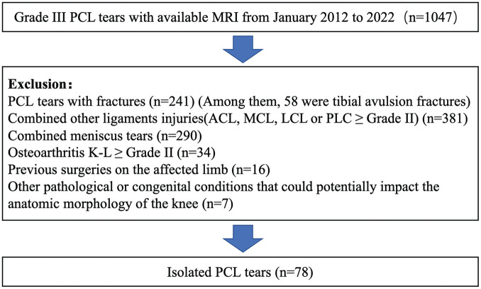
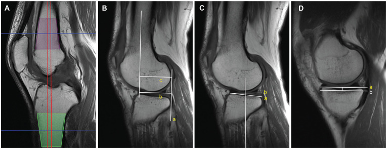
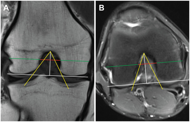
Methods: From the patients who visited the outpatient clinic of our hospital between 2012 and 2022, we included 78 patients with isolated PCL ruptures and 78 age- and sex-matched controls with knee injury but no structural damage evident on magnetic resonance imaging (MRI). The following parameters were assessed using MRI: the lateral femoral condyle and medial femoral condyle height and depth ratio (LFC-H/D, MFC-H/D), notch width index, intercondylar notch angle, the lateral tibial plateau slope, and medial tibial plateau slope (MTPS), and medial tibial depth. Values were compared between these 2 groups using the independent t test and the Mann-Whitney U test. Univariate logistic regression analysis was subsequently performed to identify independent risk factors. Receiver operating characteristic curves were generated for the morphological indicators and the combination of risk factors.
Results: Patients in the isolated PCL rupture group had significantly lower LFC-H/D (0.49 vs 0.52; P < .001) and MTPS (7.34 vs 8.81; P = .012) values compared with the control group. In sex-specific analyses, both male and female patients with isolated PCL rupture had a significantly lower LFC-H/D (female patients: 0.46 vs 0.52; P < .001; male patients: 0.51 vs 0.52; P = .035). In addition, male patients with PCL rupture had a smaller MTPS (6.74 mm vs 8.79 mm; P = .004). Univariate logistic regression analysis further validated LFC-H/D (odds ratio [OR], <0.001; P < .001) and MTPS (OR, 0.889; P = .014) as risk factors for isolated PCL rupture.
Conclusion: A decreased LFC-H/D and a reduced MTPS were identified as risk factors for isolated PCL rupture. Sex-specific analysis further suggested that a decreased LFC-H/D was a risk factor for isolated PCL ruptures in both male and female patients, whereas a lower MTPS was a risk factor exclusively in male patients.
背景:股骨髁的形态特征作为膝关节损伤的潜在危险因素最近引起了人们的关注。然而,关于股骨髁形态是否与后交叉韧带(PCL)损伤有关,我们知之甚少。目的:探讨股骨髁形态特征是否为孤立性PCL破裂的危险因素。研究设计:病例对照研究;证据水平,3。方法:选取2012年至2022年在我院门诊就诊的78例孤立性PCL破裂患者和78例年龄和性别匹配的对照组,均为膝关节损伤,但磁共振成像(MRI)未见结构损伤。MRI评估以下参数:股骨外侧髁与股骨内侧髁高度深度比(LFC-H/D, MFC-H/D)、切迹宽度指数、髁间切迹角度、胫骨外侧平台斜率、胫骨内侧平台斜率(MTPS)、胫骨内侧深度。采用独立t检验和Mann-Whitney U检验比较两组间的数值。随后进行单变量logistic回归分析以确定独立危险因素。生成形态学指标及危险因素组合的受试者工作特征曲线。结果:分离性PCL破裂组患者LFC-H/D显著降低(0.49 vs 0.52;P < 0.001)和MTPS (7.34 vs 8.81;P = 0.012)值与对照组比较。在性别特异性分析中,孤立PCL破裂的男性和女性患者的LFC-H/D均显著降低(女性患者:0.46 vs 0.52;P < .001;男性患者:0.51 vs 0.52;P = .035)。此外,男性PCL破裂患者的MTPS更小(6.74 mm vs 8.79 mm;P = .004)。单因素logistic回归分析进一步验证了LFC-H/D(比值比[OR], P < .001)和MTPS (OR, 0.889;P = 0.014)是孤立性PCL破裂的危险因素。结论:LFC-H/D降低和MTPS降低是孤立性PCL破裂的危险因素。性别特异性分析进一步表明,LFC-H/D降低是男性和女性患者孤立性PCL破裂的危险因素,而MTPS降低仅是男性患者的危险因素。
期刊介绍:
The Orthopaedic Journal of Sports Medicine (OJSM), developed by the American Orthopaedic Society for Sports Medicine (AOSSM), is a global, peer-reviewed, open access journal that combines the interests of researchers and clinical practitioners across orthopaedic sports medicine, arthroscopy, and knee arthroplasty.
Topics include original research in the areas of:
-Orthopaedic Sports Medicine, including surgical and nonsurgical treatment of orthopaedic sports injuries
-Arthroscopic Surgery (Shoulder/Elbow/Wrist/Hip/Knee/Ankle/Foot)
-Relevant translational research
-Sports traumatology/epidemiology
-Knee and shoulder arthroplasty
The OJSM also publishes relevant systematic reviews and meta-analyses.
This journal is a member of the Committee on Publication Ethics (COPE).

 求助内容:
求助内容: 应助结果提醒方式:


