{"title":"Intraosseous schwannoma of the olecranon: A case report.","authors":"Ömer Faruk Naldöven, Nevzat Arıcan, Şahan Güven, Servet Güreşci, Şahin Çepni","doi":"10.52312/jdrs.2025.2125","DOIUrl":null,"url":null,"abstract":"<p><p>Intraosseous schwannomas are extremely rare benign tumors originating from Schwann cells of the peripheral nervous system. While these tumors are commonly found in the mandible and sacrum, their occurrence in long bones, particularly the ulna, is uncommon. A 59-year-old female patient was admitted with a two-year history of pain and swelling in her right elbow. Radiographic imaging revealed a lytic lesion with cortical expansion and thinning in the proximal ulna metaphysis, while magnetic resonance imaging showed a lesion with irregular borders extending into the joint via cortical erosion. Differential diagnoses included a simple bone cyst, enchondroma, giant cell tumor, and metastasis. Surgical procedure involved curettage and bone grafting, and the lesion was diagnosed histologically as an intraosseous schwannoma. Immunohistochemical staining showed positive expression of SOX10 and S100, confirming the diagnosis. Postoperative recovery was uneventful, and the patient regained a full range of motion without pain at her six-month follow-up. In conclusion, this case underscores the importance of considering intraosseous schwannoma in the differential diagnosis of benign bone lesions, particularly in rare locations such as the ulna. Histopathological examination remains essential for an accurate diagnosis.</p>","PeriodicalId":73560,"journal":{"name":"Joint diseases and related surgery","volume":"36 3","pages":"772-777"},"PeriodicalIF":1.9000,"publicationDate":"2025-07-25","publicationTypes":"Journal Article","fieldsOfStudy":null,"isOpenAccess":false,"openAccessPdf":"https://www.ncbi.nlm.nih.gov/pmc/articles/PMC12456367/pdf/","citationCount":"0","resultStr":null,"platform":"Semanticscholar","paperid":null,"PeriodicalName":"Joint diseases and related surgery","FirstCategoryId":"1085","ListUrlMain":"https://doi.org/10.52312/jdrs.2025.2125","RegionNum":0,"RegionCategory":null,"ArticlePicture":[],"TitleCN":null,"AbstractTextCN":null,"PMCID":null,"EPubDate":"","PubModel":"","JCR":"Q2","JCRName":"ORTHOPEDICS","Score":null,"Total":0}
引用次数: 0
Abstract
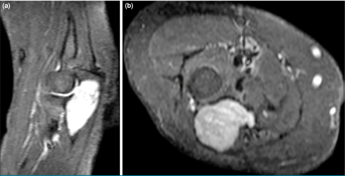
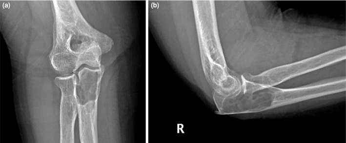
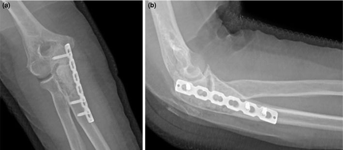
Intraosseous schwannomas are extremely rare benign tumors originating from Schwann cells of the peripheral nervous system. While these tumors are commonly found in the mandible and sacrum, their occurrence in long bones, particularly the ulna, is uncommon. A 59-year-old female patient was admitted with a two-year history of pain and swelling in her right elbow. Radiographic imaging revealed a lytic lesion with cortical expansion and thinning in the proximal ulna metaphysis, while magnetic resonance imaging showed a lesion with irregular borders extending into the joint via cortical erosion. Differential diagnoses included a simple bone cyst, enchondroma, giant cell tumor, and metastasis. Surgical procedure involved curettage and bone grafting, and the lesion was diagnosed histologically as an intraosseous schwannoma. Immunohistochemical staining showed positive expression of SOX10 and S100, confirming the diagnosis. Postoperative recovery was uneventful, and the patient regained a full range of motion without pain at her six-month follow-up. In conclusion, this case underscores the importance of considering intraosseous schwannoma in the differential diagnosis of benign bone lesions, particularly in rare locations such as the ulna. Histopathological examination remains essential for an accurate diagnosis.

 求助内容:
求助内容: 应助结果提醒方式:


