{"title":"Evaluation of delayed diagnosis of osteoid osteoma in adolescents and young patients with hip pain.","authors":"Ferit Tufan Özgezmez, Gül Öznur Karabıçak, Emre Çullu","doi":"10.52312/jdrs.2025.2199","DOIUrl":null,"url":null,"abstract":"<p><strong>Objectives: </strong>This study aims to evaluate delayed diagnosis of osteoid osteoma (OO) in pediatric patients with hip pain and to identify the diagnostic challenges and errors encountered during this period.</p><p><strong>Patients and methods: </strong>Between May 2010 and July 2022, a total of 18 patients (11 males; 7 females; median age: 12 years; range, 6 to 20 years) with a confirmed diagnosis of OO were retrospectively analyzed. Demographic, clinical, and radiographic data were recorded. The time from symptom onset to diagnosis was calculated for each patient. The date of radiological diagnosis was accepted as the time of diagnosis.</p><p><strong>Results: </strong>The median time from symptom onset to diagnosis was 12 months. Right-sided involvement was observed in 61.1% of cases. The most common lesion location was the femoral neck (61.1%), and 66.7% of cases had intra-articular lesions. A limping gait was observed in 61.1% of patients. Additionally, 33.3% of cases reported atrophy of the thigh muscles and/or lower extremities. Night pain was present in 83.3% of cases. A total of 72.2% of cases had a diagnostic delay exceeding six months. Half (50%) of the patients required more than five visits to healthcare providers before receiving an accurate diagnosis.</p><p><strong>Conclusion: </strong>The diagnostic delays for OO located in the hip region can be seen in among children and young adults, primarily due to misdiagnosis and reliance on inconclusive initial imaging findings. To minimize such delays, clinicians should maintain a high index of suspicion, particularly in patients with persistent, unexplained hip pain, and consider imaging studies. Pain lasting over three weeks warrants further diagnostic evaluation in this patient group.</p>","PeriodicalId":73560,"journal":{"name":"Joint diseases and related surgery","volume":"36 3","pages":"640-647"},"PeriodicalIF":1.9000,"publicationDate":"2025-07-07","publicationTypes":"Journal Article","fieldsOfStudy":null,"isOpenAccess":false,"openAccessPdf":"https://www.ncbi.nlm.nih.gov/pmc/articles/PMC12456355/pdf/","citationCount":"0","resultStr":null,"platform":"Semanticscholar","paperid":null,"PeriodicalName":"Joint diseases and related surgery","FirstCategoryId":"1085","ListUrlMain":"https://doi.org/10.52312/jdrs.2025.2199","RegionNum":0,"RegionCategory":null,"ArticlePicture":[],"TitleCN":null,"AbstractTextCN":null,"PMCID":null,"EPubDate":"","PubModel":"","JCR":"Q2","JCRName":"ORTHOPEDICS","Score":null,"Total":0}
引用次数: 0
Abstract
Objectives: This study aims to evaluate delayed diagnosis of osteoid osteoma (OO) in pediatric patients with hip pain and to identify the diagnostic challenges and errors encountered during this period.
Patients and methods: Between May 2010 and July 2022, a total of 18 patients (11 males; 7 females; median age: 12 years; range, 6 to 20 years) with a confirmed diagnosis of OO were retrospectively analyzed. Demographic, clinical, and radiographic data were recorded. The time from symptom onset to diagnosis was calculated for each patient. The date of radiological diagnosis was accepted as the time of diagnosis.
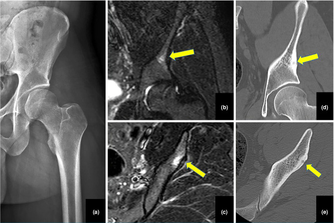
Results: The median time from symptom onset to diagnosis was 12 months. Right-sided involvement was observed in 61.1% of cases. The most common lesion location was the femoral neck (61.1%), and 66.7% of cases had intra-articular lesions. A limping gait was observed in 61.1% of patients. Additionally, 33.3% of cases reported atrophy of the thigh muscles and/or lower extremities. Night pain was present in 83.3% of cases. A total of 72.2% of cases had a diagnostic delay exceeding six months. Half (50%) of the patients required more than five visits to healthcare providers before receiving an accurate diagnosis.
Conclusion: The diagnostic delays for OO located in the hip region can be seen in among children and young adults, primarily due to misdiagnosis and reliance on inconclusive initial imaging findings. To minimize such delays, clinicians should maintain a high index of suspicion, particularly in patients with persistent, unexplained hip pain, and consider imaging studies. Pain lasting over three weeks warrants further diagnostic evaluation in this patient group.

 求助内容:
求助内容: 应助结果提醒方式:


