Turkay Yilmaz, Ismail Hakki Dur, Tugce Kabakci, Muhammed Abdulkadir Bulut, Bengu Akgok, Ulas Can Kolac, Mustafa Ozkaya, Sancar Bakircioglu
{"title":"Effect of fracture level on optimal Kirschner wire configuration in pediatric supracondylar humerus fractures: A finite element analysis.","authors":"Turkay Yilmaz, Ismail Hakki Dur, Tugce Kabakci, Muhammed Abdulkadir Bulut, Bengu Akgok, Ulas Can Kolac, Mustafa Ozkaya, Sancar Bakircioglu","doi":"10.52312/jdrs.2025.2248","DOIUrl":null,"url":null,"abstract":"<p><strong>Objectives: </strong>This study aims to evaluate the biomechanical stability of three pin configurations for transverse supracondylar humerus fractures at various levels using finite element analysis (FEA).</p><p><strong>Materials and methods: </strong>Computed tomography data from a six-year-old child were used to generate a humerus bone model. Four different fracture levels (low, transolecranon, high, and ultrahigh) and three pin fixation techniques (one lateral and one medial cross-pin [1-1M], two lateral capitellar pins [1-1C], and three lateral capitellar pins [2-1C]) were designed for the study. Translational stiffness and rotational stiffness in all directions were analyzed in the mesh models. Convergence data and stiffness data were obtained in the FEA.</p><p><strong>Results: </strong>The translational and rotational stiffness values varied across fracture levels and pin configurations. Under valgus loading, the 1-1M configuration provided the highest stability in ultrahigh fractures (3289 N/mm), while the 2-1C configuration showed superior valgus and varus stability in low and transolecranon fractures. During extension and flexion loading, the 1-1M configuration yielded the highest stiffness values for transolecranon and high fractures, while the 2-1C configuration demonstrated increased stability in low and ultrahigh fractures. For rotational loading, 1-1M produced the highest inward and outward stiffness values in low-level fractures (9175 and 11035 N·mm/degree, respectively), whereas 2-1C displayed greater rotational stiffness in ultrahigh fractures.</p><p><strong>Conclusion: </strong>This preliminary study suggests that no single pin configuration is ideal for all fracture types, and the choice should be based on the specific fracture case.</p>","PeriodicalId":73560,"journal":{"name":"Joint diseases and related surgery","volume":"36 3","pages":"648-658"},"PeriodicalIF":1.9000,"publicationDate":"2025-07-21","publicationTypes":"Journal Article","fieldsOfStudy":null,"isOpenAccess":false,"openAccessPdf":"https://www.ncbi.nlm.nih.gov/pmc/articles/PMC12456352/pdf/","citationCount":"0","resultStr":null,"platform":"Semanticscholar","paperid":null,"PeriodicalName":"Joint diseases and related surgery","FirstCategoryId":"1085","ListUrlMain":"https://doi.org/10.52312/jdrs.2025.2248","RegionNum":0,"RegionCategory":null,"ArticlePicture":[],"TitleCN":null,"AbstractTextCN":null,"PMCID":null,"EPubDate":"","PubModel":"","JCR":"Q2","JCRName":"ORTHOPEDICS","Score":null,"Total":0}
引用次数: 0
Abstract
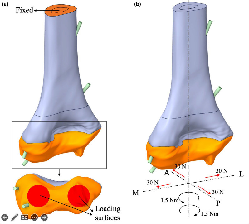
Objectives: This study aims to evaluate the biomechanical stability of three pin configurations for transverse supracondylar humerus fractures at various levels using finite element analysis (FEA).
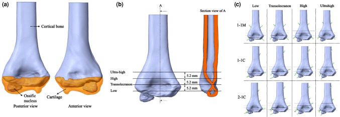
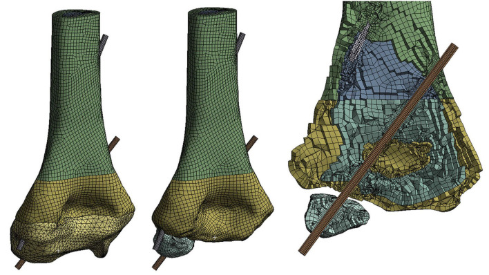
Materials and methods: Computed tomography data from a six-year-old child were used to generate a humerus bone model. Four different fracture levels (low, transolecranon, high, and ultrahigh) and three pin fixation techniques (one lateral and one medial cross-pin [1-1M], two lateral capitellar pins [1-1C], and three lateral capitellar pins [2-1C]) were designed for the study. Translational stiffness and rotational stiffness in all directions were analyzed in the mesh models. Convergence data and stiffness data were obtained in the FEA.
Results: The translational and rotational stiffness values varied across fracture levels and pin configurations. Under valgus loading, the 1-1M configuration provided the highest stability in ultrahigh fractures (3289 N/mm), while the 2-1C configuration showed superior valgus and varus stability in low and transolecranon fractures. During extension and flexion loading, the 1-1M configuration yielded the highest stiffness values for transolecranon and high fractures, while the 2-1C configuration demonstrated increased stability in low and ultrahigh fractures. For rotational loading, 1-1M produced the highest inward and outward stiffness values in low-level fractures (9175 and 11035 N·mm/degree, respectively), whereas 2-1C displayed greater rotational stiffness in ultrahigh fractures.
Conclusion: This preliminary study suggests that no single pin configuration is ideal for all fracture types, and the choice should be based on the specific fracture case.

 求助内容:
求助内容: 应助结果提醒方式:


