Ugur Dalaman, Ibrahim Cuneyit, Şevval Öztürk, Ege Rıza Karagur, Mert Ocak, Nazmi Yaras, Mustafa Unal, Baris Ozgur Donmez
{"title":"Potential preventive effects of angiotensin-(1-7) on bone matrix quality in diabetic rats through modulation of the organic matrix.","authors":"Ugur Dalaman, Ibrahim Cuneyit, Şevval Öztürk, Ege Rıza Karagur, Mert Ocak, Nazmi Yaras, Mustafa Unal, Baris Ozgur Donmez","doi":"10.52312/jdrs.2025.2181","DOIUrl":null,"url":null,"abstract":"<p><strong>Objectives: </strong>This experimental study aims to investigate the effects of angiotensin (1-7) (Ang-[1-7]) on the microstructural, biomechanical, and biophysicochemical properties of bone tissue in diabetic rats.</p><p><strong>Materials and methods: </strong>Forty-eight male Wistar rats, aged three months and weighing between 280 and 330 g, were used in this study. Four groups, each containing 12 rats, were established: Control, diabetes mellitus (DM), DM-Ang-(1-7), and Ang-(1-7). The samples underwent analysis through micro-computed tomography (CT), Raman spectroscopy, and three-point bending biomechanical test.</p><p><strong>Results: </strong>Diabetes significantly impaired bone quality, with reduced cortical thickness, maximum load, and flexural strength (p<0.05). The Ang-(1-7) treatment improved flexural strength (p<0.05), but did not fully restore mechanical function. Micro-CT showed decreased bone volume and trabecular thickness in both diabetic groups (p<0.05), with no significant recovery by Ang-(1-7). Raman spectroscopy revealed lower mineral-to-matrix ratio and disrupted collagen quality in diabetic bone (p<0.05), while Ang-(1-7) partially restored collagen-related parameters.</p><p><strong>Conclusion: </strong>These findings highlight that Ang-(1-7) has minimal impact on bone minerals in DM rats. However, it may have a potential preventive effect on the triple-helix structural impairment within the bone organic matrix in this model.</p>","PeriodicalId":73560,"journal":{"name":"Joint diseases and related surgery","volume":"36 3","pages":"577-588"},"PeriodicalIF":1.9000,"publicationDate":"2025-07-21","publicationTypes":"Journal Article","fieldsOfStudy":null,"isOpenAccess":false,"openAccessPdf":"https://www.ncbi.nlm.nih.gov/pmc/articles/PMC12456337/pdf/","citationCount":"0","resultStr":null,"platform":"Semanticscholar","paperid":null,"PeriodicalName":"Joint diseases and related surgery","FirstCategoryId":"1085","ListUrlMain":"https://doi.org/10.52312/jdrs.2025.2181","RegionNum":0,"RegionCategory":null,"ArticlePicture":[],"TitleCN":null,"AbstractTextCN":null,"PMCID":null,"EPubDate":"","PubModel":"","JCR":"Q2","JCRName":"ORTHOPEDICS","Score":null,"Total":0}
引用次数: 0
Abstract
Objectives: This experimental study aims to investigate the effects of angiotensin (1-7) (Ang-[1-7]) on the microstructural, biomechanical, and biophysicochemical properties of bone tissue in diabetic rats.
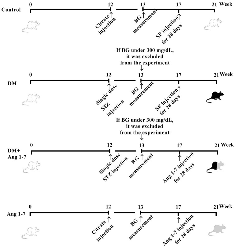
Materials and methods: Forty-eight male Wistar rats, aged three months and weighing between 280 and 330 g, were used in this study. Four groups, each containing 12 rats, were established: Control, diabetes mellitus (DM), DM-Ang-(1-7), and Ang-(1-7). The samples underwent analysis through micro-computed tomography (CT), Raman spectroscopy, and three-point bending biomechanical test.
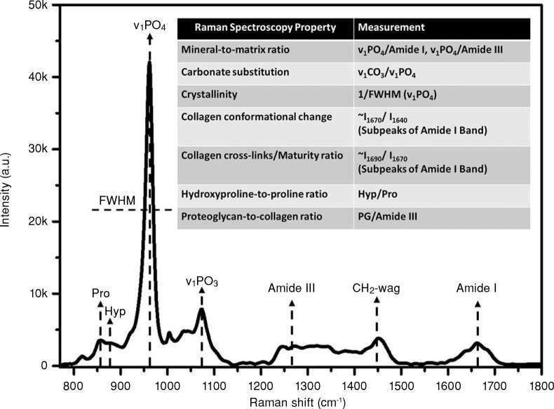
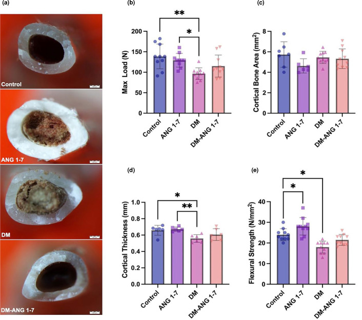
Results: Diabetes significantly impaired bone quality, with reduced cortical thickness, maximum load, and flexural strength (p<0.05). The Ang-(1-7) treatment improved flexural strength (p<0.05), but did not fully restore mechanical function. Micro-CT showed decreased bone volume and trabecular thickness in both diabetic groups (p<0.05), with no significant recovery by Ang-(1-7). Raman spectroscopy revealed lower mineral-to-matrix ratio and disrupted collagen quality in diabetic bone (p<0.05), while Ang-(1-7) partially restored collagen-related parameters.
Conclusion: These findings highlight that Ang-(1-7) has minimal impact on bone minerals in DM rats. However, it may have a potential preventive effect on the triple-helix structural impairment within the bone organic matrix in this model.

 求助内容:
求助内容: 应助结果提醒方式:


