Meningeal lymphatic dysfunction in sporadic cerebral small vessel diseases and cerebral autosomal dominant arteriopathy with subcortical infarcts and leukoencephalopathy by DCE-MRI.
IF 2.3 2区 医学Q2 RADIOLOGY, NUCLEAR MEDICINE & MEDICAL IMAGING
Han Wang, Xiao Ren, Yu Shen, Yusen Qiu, Menghua Li, Si Luo, Zhi Zhang, Lin Wu, Meihong Zhou, Yixin Jiang, Fuqing Zhou, Daojun Hong
{"title":"Meningeal lymphatic dysfunction in sporadic cerebral small vessel diseases and cerebral autosomal dominant arteriopathy with subcortical infarcts and leukoencephalopathy by DCE-MRI.","authors":"Han Wang, Xiao Ren, Yu Shen, Yusen Qiu, Menghua Li, Si Luo, Zhi Zhang, Lin Wu, Meihong Zhou, Yixin Jiang, Fuqing Zhou, Daojun Hong","doi":"10.21037/qims-24-2429","DOIUrl":null,"url":null,"abstract":"<p><strong>Background: </strong>The distinction between cerebral autosomal dominant arteriopathy with subcortical infarcts and leukoencephalopathy (CADASIL), a prevalent hereditary cerebrovascular disease, and sporadic cerebrovascular disease has sparked significant interest among researchers. Meningeal lymphatic vessels (mLVs) have become a new research topic in recent years. This study aimed to investigate the function of meningeal lymphatics in cerebral small vessel disease (CSVD) and to develop a predictive model to assist in the diagnosis of CADASIL patients.</p><p><strong>Methods: </strong>We systematically examined the functional changes in the meningeal lymphatic system in both sporadic CSVD patients and CADASIL patients using dynamic contrast-enhanced magnetic resonance imaging (DCE-MRI), and the correlations among blood biomarkers were subsequently calculated. Finally, a nomogram was generated for the identification of CADASIL using multiple significant risk factors.</p><p><strong>Results: </strong>This study enrolled 41 sporadic CSVD (S-CSVD) patients, 15 CADASIL patients, and 18 normal controls (NC). Both CSVD and CADASIL groups demonstrated mLVs dysfunction compared to controls, characterized by decreased mLVs wash-in rates (P<0.05) and prolonged time-to-peak (TTP, P<0.01). Significant metabolic correlations emerged specifically in CSVD: In the S-CSVD group, the parameters of DCE-MRI [wash-in rate, TTP, incremental area under the curve (IAUC), transfer rate constant (Ktrans), extravascular extracellular volume fraction (Ve)] correlated with total cholesterol (TC, TTP, r=-0.38), low-density lipoprotein cholesterol (LDL-C, TTP, r=-0.41), serum creatinine (SCr, Ktrans, r=0.34), uric acid (UA, Ve, r=0.34), homocysteine (Hcy, wash-in, r=-0.31, both P<0.05), and hemoglobin A1c (HbA1c, wash-in; r=-0.32, P<0.01, IAUC; r=-0.47, P<0.01). In the CADASIL group, the parameter of DCE-MRI correlated with folic acid (FA, TTP, r=-0.57, P=0.03), SCr (IAUC; r=0.54, P=0.04, Ktrans; r=0.45, P=0.01, Ve; r=0.61, P<0.01), and UA (Ktrans; r=0.45, Ve; r=0.71, both P<0.05). Additionally, we identified several critical risk factors for diagnosing CADASIL and developed a nomogram to differentiate CADASIL from S-CSVD (area under the curve 0.870, 95% confidence interval: 0.767-0.973).</p><p><strong>Conclusions: </strong>This study confirmed that mLVs were dysfunctional in both S-CSVD patients and CADASIL patients. IAUC was identified as a key risk factor, and a nomogram was generated. This research enhances our understanding of the role of mLVs in CSVD and contributes to distinguishing between these S-CSVD and CADASIL.</p>","PeriodicalId":54267,"journal":{"name":"Quantitative Imaging in Medicine and Surgery","volume":"15 8","pages":"6692-6704"},"PeriodicalIF":2.3000,"publicationDate":"2025-08-01","publicationTypes":"Journal Article","fieldsOfStudy":null,"isOpenAccess":false,"openAccessPdf":"https://www.ncbi.nlm.nih.gov/pmc/articles/PMC12332741/pdf/","citationCount":"0","resultStr":null,"platform":"Semanticscholar","paperid":null,"PeriodicalName":"Quantitative Imaging in Medicine and Surgery","FirstCategoryId":"3","ListUrlMain":"https://doi.org/10.21037/qims-24-2429","RegionNum":2,"RegionCategory":"医学","ArticlePicture":[],"TitleCN":null,"AbstractTextCN":null,"PMCID":null,"EPubDate":"2025/7/28 0:00:00","PubModel":"Epub","JCR":"Q2","JCRName":"RADIOLOGY, NUCLEAR MEDICINE & MEDICAL IMAGING","Score":null,"Total":0}
引用次数: 0
Abstract
Background: The distinction between cerebral autosomal dominant arteriopathy with subcortical infarcts and leukoencephalopathy (CADASIL), a prevalent hereditary cerebrovascular disease, and sporadic cerebrovascular disease has sparked significant interest among researchers. Meningeal lymphatic vessels (mLVs) have become a new research topic in recent years. This study aimed to investigate the function of meningeal lymphatics in cerebral small vessel disease (CSVD) and to develop a predictive model to assist in the diagnosis of CADASIL patients.
Methods: We systematically examined the functional changes in the meningeal lymphatic system in both sporadic CSVD patients and CADASIL patients using dynamic contrast-enhanced magnetic resonance imaging (DCE-MRI), and the correlations among blood biomarkers were subsequently calculated. Finally, a nomogram was generated for the identification of CADASIL using multiple significant risk factors.
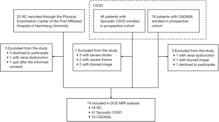
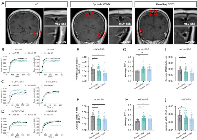
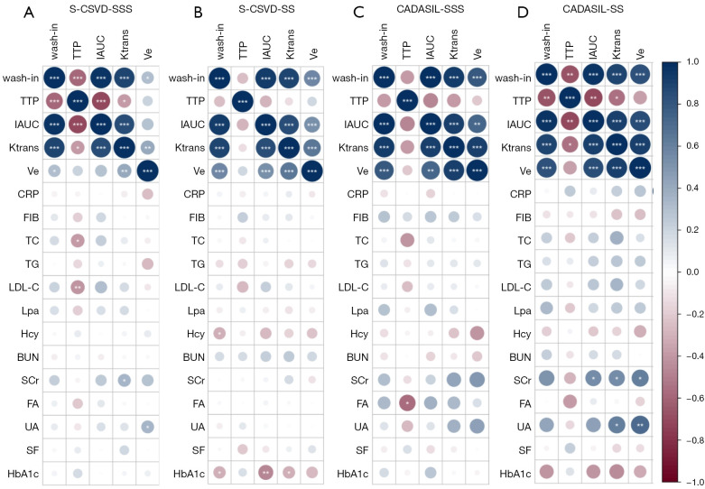
Results: This study enrolled 41 sporadic CSVD (S-CSVD) patients, 15 CADASIL patients, and 18 normal controls (NC). Both CSVD and CADASIL groups demonstrated mLVs dysfunction compared to controls, characterized by decreased mLVs wash-in rates (P<0.05) and prolonged time-to-peak (TTP, P<0.01). Significant metabolic correlations emerged specifically in CSVD: In the S-CSVD group, the parameters of DCE-MRI [wash-in rate, TTP, incremental area under the curve (IAUC), transfer rate constant (Ktrans), extravascular extracellular volume fraction (Ve)] correlated with total cholesterol (TC, TTP, r=-0.38), low-density lipoprotein cholesterol (LDL-C, TTP, r=-0.41), serum creatinine (SCr, Ktrans, r=0.34), uric acid (UA, Ve, r=0.34), homocysteine (Hcy, wash-in, r=-0.31, both P<0.05), and hemoglobin A1c (HbA1c, wash-in; r=-0.32, P<0.01, IAUC; r=-0.47, P<0.01). In the CADASIL group, the parameter of DCE-MRI correlated with folic acid (FA, TTP, r=-0.57, P=0.03), SCr (IAUC; r=0.54, P=0.04, Ktrans; r=0.45, P=0.01, Ve; r=0.61, P<0.01), and UA (Ktrans; r=0.45, Ve; r=0.71, both P<0.05). Additionally, we identified several critical risk factors for diagnosing CADASIL and developed a nomogram to differentiate CADASIL from S-CSVD (area under the curve 0.870, 95% confidence interval: 0.767-0.973).
Conclusions: This study confirmed that mLVs were dysfunctional in both S-CSVD patients and CADASIL patients. IAUC was identified as a key risk factor, and a nomogram was generated. This research enhances our understanding of the role of mLVs in CSVD and contributes to distinguishing between these S-CSVD and CADASIL.

 求助内容:
求助内容: 应助结果提醒方式:


