Anatomical Pelvic Parameters Using the Anterior Pelvic Plane: Relationships with Standing Sagittal Spinal Alignment and Estimated Lumbar Alignment in Healthy Volunteers.
{"title":"Anatomical Pelvic Parameters Using the Anterior Pelvic Plane: Relationships with Standing Sagittal Spinal Alignment and Estimated Lumbar Alignment in Healthy Volunteers.","authors":"Masayuki Ohashi, Kazuhiro Hasegawa, Shun Hatsushikano, Norio Imai, Hideki Tashi, Yohei Shibuya, Keitaro Minato, Masayuki Sato, Hiroyuki Sekimoto, Kei Watanabe, Hiroyuki Kawashima","doi":"10.22603/ssrr.2024-0283","DOIUrl":null,"url":null,"abstract":"<p><strong>Introduction: </strong>To estimate natural standing sagittal alignment in patients with adult spinal deformity (ASD), we previously reported the normative values of anatomical pelvic parameters in a healthy population, based on the anterior pelvic plane (APP), and observed the relationships between anatomical and positional pelvic parameters in the standing position. As the second step, we aim to investigate the relationships between anatomical pelvic parameters and standing spinal sagittal alignment in a healthy population.</p><p><strong>Methods: </strong>We analyzed biplanar, slot-scanning, full-body stereo radiography of 140 healthy Japanese volunteers (mean age, 39.5 years; 59.3% women). The APP was defined by bilateral anterior superior iliac spines and anterior surface of the pubis symphysis. Anatomical sacral slope (aSS) and anatomical pelvic tilt (aPT) were calculated as angles of the SS and PT regarding the APP.</p><p><strong>Results: </strong>The APP was tilted anteriorly in the sagittal plane by an average of 0.7°. Anatomical pelvic parameters significantly correlated with standing sagittal parameters, except for cervical lordosis and T4-12 thoracic kyphosis (TK) (p<0.05). L4-S1 lumbar lordosis (LL) significantly correlated with aPT and aSS, but not with pelvic incidence (PI). In addition, T1-12 TK significantly correlated with aSS. Multiple linear regression analysis for lumbar alignment produced the following equations: L1-S1 LL (°)=0.588×aSS+30.522, L4-S1 LL (°)=0.165×aSS-0.248×aPT+32.825, lordosis distribution index (%)=-0.662×PI+102.8.</p><p><strong>Conclusions: </strong>Novel relationships in a healthy population were identified between the anatomical characteristics of the pelvis and standing sagittal parameters not represented by PI. This novel measurement concept based on the APP may estimate natural standing sagittal alignments and proportions using anatomical pelvic parameters in ASD.</p>","PeriodicalId":22253,"journal":{"name":"Spine Surgery and Related Research","volume":"9 4","pages":"469-476"},"PeriodicalIF":1.2000,"publicationDate":"2024-12-20","publicationTypes":"Journal Article","fieldsOfStudy":null,"isOpenAccess":false,"openAccessPdf":"https://www.ncbi.nlm.nih.gov/pmc/articles/PMC12330368/pdf/","citationCount":"0","resultStr":null,"platform":"Semanticscholar","paperid":null,"PeriodicalName":"Spine Surgery and Related Research","FirstCategoryId":"1085","ListUrlMain":"https://doi.org/10.22603/ssrr.2024-0283","RegionNum":0,"RegionCategory":null,"ArticlePicture":[],"TitleCN":null,"AbstractTextCN":null,"PMCID":null,"EPubDate":"2025/7/27 0:00:00","PubModel":"eCollection","JCR":"Q3","JCRName":"SURGERY","Score":null,"Total":0}
引用次数: 0
Abstract
Introduction: To estimate natural standing sagittal alignment in patients with adult spinal deformity (ASD), we previously reported the normative values of anatomical pelvic parameters in a healthy population, based on the anterior pelvic plane (APP), and observed the relationships between anatomical and positional pelvic parameters in the standing position. As the second step, we aim to investigate the relationships between anatomical pelvic parameters and standing spinal sagittal alignment in a healthy population.
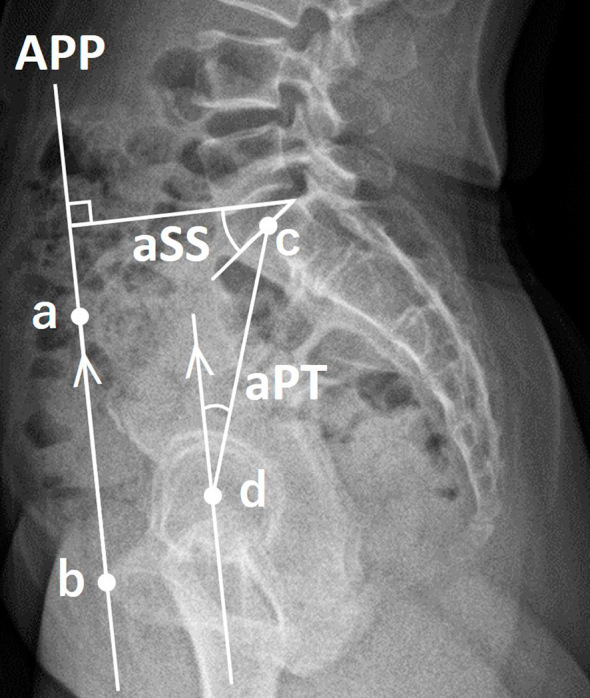
Methods: We analyzed biplanar, slot-scanning, full-body stereo radiography of 140 healthy Japanese volunteers (mean age, 39.5 years; 59.3% women). The APP was defined by bilateral anterior superior iliac spines and anterior surface of the pubis symphysis. Anatomical sacral slope (aSS) and anatomical pelvic tilt (aPT) were calculated as angles of the SS and PT regarding the APP.
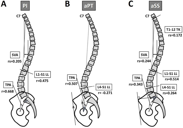
Results: The APP was tilted anteriorly in the sagittal plane by an average of 0.7°. Anatomical pelvic parameters significantly correlated with standing sagittal parameters, except for cervical lordosis and T4-12 thoracic kyphosis (TK) (p<0.05). L4-S1 lumbar lordosis (LL) significantly correlated with aPT and aSS, but not with pelvic incidence (PI). In addition, T1-12 TK significantly correlated with aSS. Multiple linear regression analysis for lumbar alignment produced the following equations: L1-S1 LL (°)=0.588×aSS+30.522, L4-S1 LL (°)=0.165×aSS-0.248×aPT+32.825, lordosis distribution index (%)=-0.662×PI+102.8.
Conclusions: Novel relationships in a healthy population were identified between the anatomical characteristics of the pelvis and standing sagittal parameters not represented by PI. This novel measurement concept based on the APP may estimate natural standing sagittal alignments and proportions using anatomical pelvic parameters in ASD.

 求助内容:
求助内容: 应助结果提醒方式:


