{"title":"Abnormal Paravertebral Muscle Activity and Cervical Extensor Muscle Condition Affect Dynamic Spinal Balance during Gait in Dropped Head Syndrome.","authors":"Kotaro Sakashita, Kousei Miura, Hideki Kadone, Tomoyuki Asada, Takahiro Sunami, Takane Nakagawa, Yosuke Ogata, Shun Okuwaki, Tomoaki Shimizu, Hisanori Gamada, Hiroshi Noguchi, Hiroshi Takahashi, Toru Funayama, Masao Koda, Masashi Yamazaki","doi":"10.22603/ssrr.2024-0263","DOIUrl":null,"url":null,"abstract":"<p><strong>Introduction: </strong>The pathogenesis of dropped head syndrome (DHS) involves factors like fat infiltration of the cervical extensor muscle, cervical degeneration, and sarcopenia, which are typically assessed using conventional imaging. Previous studies have demonstrated cervical and thoracic anterior tilt deterioration during gait in patients with DHS. However, the relationship between dynamic spinal balance and conventional imaging findings has not been investigated. The purpose of this study was to investigate the walking posture of patients with DHS using 3D gait motion analysis and to analyze the relationship between dynamic posture and conventional imaging factors, leading to the investigation of the pathophysiology of cervical imbalance during gait in patients with DHS.</p><p><strong>Methods: </strong>Twenty-two patients with DHS were included. Global and cervical static alignments were assessed using whole spine radiography. 3D gait motion analysis was performed, and dynamic kinematic variables were segmented into the cervical and thoracic regions. The paraspinal muscle activity was assessed using wireless surface electromyography. The cervical deep extensor muscle (C-DEM) condition was assessed using magnetic resonance imaging. Correlations of changes in dynamic kinematic variables with paraspinal muscle activity and C-DEM condition were determined.</p><p><strong>Results: </strong>A significant change in the anterior cervical and thoracic spine tilt was observed during gait. These changes were inversely correlated with thoracic paraspinal muscle activity. The change in the cervical anterior tilt was significantly correlated with the fat-free C-DEM at C3/C4 and C4/C5 and the fat infiltration rate of the C-DEM at C5/C6 and C7/T1.</p><p><strong>Conclusions: </strong>The thoracic paraspinal muscle activity failed to respond to the deterioration of the thoracic anterior tilt, indicating a notable contribution to postural endurance during gait and to DHS pathogenesis. Evaluating the condition of the C-DEM could be an alternative for evaluating dynamic postural endurance and is clinically important when considering patient complaints regarding difficulties in daily activities.</p>","PeriodicalId":22253,"journal":{"name":"Spine Surgery and Related Research","volume":"9 4","pages":"398-406"},"PeriodicalIF":1.2000,"publicationDate":"2025-02-07","publicationTypes":"Journal Article","fieldsOfStudy":null,"isOpenAccess":false,"openAccessPdf":"https://www.ncbi.nlm.nih.gov/pmc/articles/PMC12330373/pdf/","citationCount":"0","resultStr":null,"platform":"Semanticscholar","paperid":null,"PeriodicalName":"Spine Surgery and Related Research","FirstCategoryId":"1085","ListUrlMain":"https://doi.org/10.22603/ssrr.2024-0263","RegionNum":0,"RegionCategory":null,"ArticlePicture":[],"TitleCN":null,"AbstractTextCN":null,"PMCID":null,"EPubDate":"2025/7/27 0:00:00","PubModel":"eCollection","JCR":"Q3","JCRName":"SURGERY","Score":null,"Total":0}
引用次数: 0
Abstract
Introduction: The pathogenesis of dropped head syndrome (DHS) involves factors like fat infiltration of the cervical extensor muscle, cervical degeneration, and sarcopenia, which are typically assessed using conventional imaging. Previous studies have demonstrated cervical and thoracic anterior tilt deterioration during gait in patients with DHS. However, the relationship between dynamic spinal balance and conventional imaging findings has not been investigated. The purpose of this study was to investigate the walking posture of patients with DHS using 3D gait motion analysis and to analyze the relationship between dynamic posture and conventional imaging factors, leading to the investigation of the pathophysiology of cervical imbalance during gait in patients with DHS.
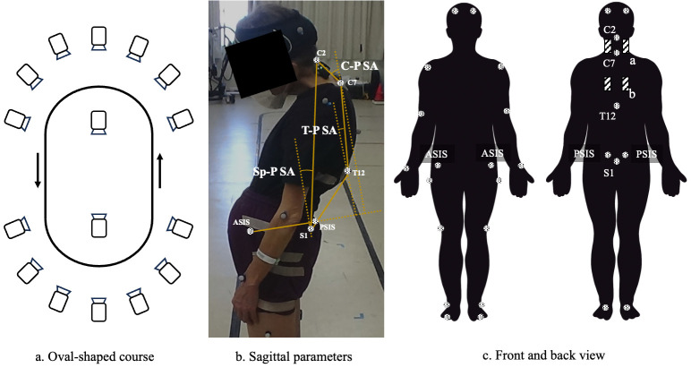
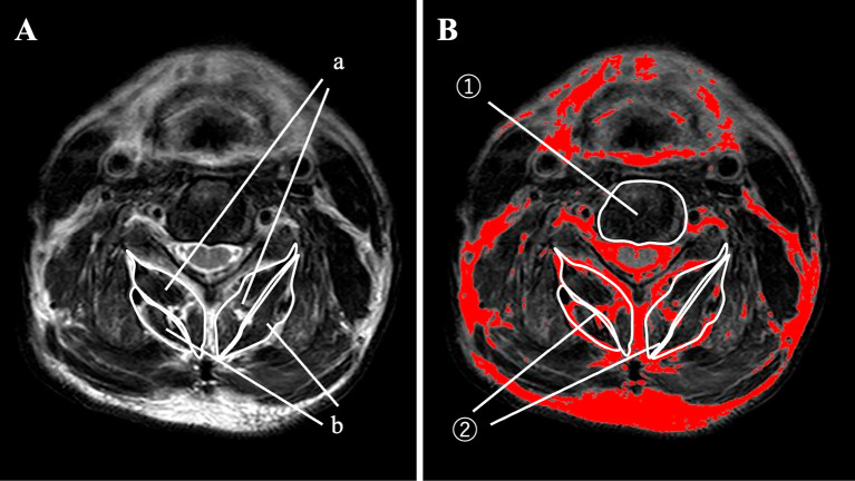
Methods: Twenty-two patients with DHS were included. Global and cervical static alignments were assessed using whole spine radiography. 3D gait motion analysis was performed, and dynamic kinematic variables were segmented into the cervical and thoracic regions. The paraspinal muscle activity was assessed using wireless surface electromyography. The cervical deep extensor muscle (C-DEM) condition was assessed using magnetic resonance imaging. Correlations of changes in dynamic kinematic variables with paraspinal muscle activity and C-DEM condition were determined.
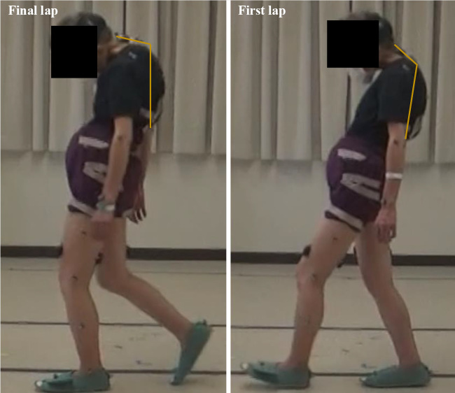
Results: A significant change in the anterior cervical and thoracic spine tilt was observed during gait. These changes were inversely correlated with thoracic paraspinal muscle activity. The change in the cervical anterior tilt was significantly correlated with the fat-free C-DEM at C3/C4 and C4/C5 and the fat infiltration rate of the C-DEM at C5/C6 and C7/T1.
Conclusions: The thoracic paraspinal muscle activity failed to respond to the deterioration of the thoracic anterior tilt, indicating a notable contribution to postural endurance during gait and to DHS pathogenesis. Evaluating the condition of the C-DEM could be an alternative for evaluating dynamic postural endurance and is clinically important when considering patient complaints regarding difficulties in daily activities.

 求助内容:
求助内容: 应助结果提醒方式:


