Chronic Syndesmotic Instability Associated with a Complex Lesion of the Posterior Inferior Tibiofibular Ligament: Review, Case Report, and Surgical Report.
{"title":"Chronic Syndesmotic Instability Associated with a Complex Lesion of the Posterior Inferior Tibiofibular Ligament: Review, Case Report, and Surgical Report.","authors":"Virginie Perez, Nermine Habib, Angela Seidel","doi":"10.13107/jocr.2025.v15.i08.5960","DOIUrl":null,"url":null,"abstract":"<p><strong>Introduction: </strong>Syndesmotic injuries, particularly those involving the posterior inferior tibiofibular ligament (PITFL), are complex and often result in chronic pain and instability if not appropriately treated. The PITFL plays a crucial role in maintaining syndesmotic stability, especially in resisting rotational forces. This case report examines a PITFL injury involving two posterior fibular fragments, supporting the hypothesis that the superficial and deep components of the ligament function independently.</p><p><strong>Case report: </strong>A 41-year-old male presented after a bicycle accident with a complex ankle fracture involving a transverse medial malleolus fracture, a postero-medial tibial fragment, a fibular tip fracture, and two additional posterior fibular fragments. Despite initial fracture management, including closed reduction and open fixation, the patient developed chronic pain and instability due to malreduction. Computed tomography imaging revealed instability of the fibula within the fibular notch, indicating syndesmotic instability. The surgical procedure included fibular osteotomy, temporary fixation with K-wires, syndesmotic fixation with the TightRope® system, and PITFL repair using the InternalBrace™ ligament augmentation system. Intraoperative three-dimensional imaging confirmed successful reduction and stabilization.</p><p><strong>Conclusion: </strong>Fibular avulsion of the PITFL is rare. Failure to diagnose the lesion may lead to malreduction of the fibula within the incisura. The combination of osteotomy, TightRope® syndesmosis fixation, and InternalBrace™ PITFL repair provides a reliable option for managing complex PITFL injuries.</p>","PeriodicalId":16647,"journal":{"name":"Journal of Orthopaedic Case Reports","volume":"15 8","pages":"256-259"},"PeriodicalIF":0.0000,"publicationDate":"2025-08-01","publicationTypes":"Journal Article","fieldsOfStudy":null,"isOpenAccess":false,"openAccessPdf":"https://www.ncbi.nlm.nih.gov/pmc/articles/PMC12328971/pdf/","citationCount":"0","resultStr":null,"platform":"Semanticscholar","paperid":null,"PeriodicalName":"Journal of Orthopaedic Case Reports","FirstCategoryId":"1085","ListUrlMain":"https://doi.org/10.13107/jocr.2025.v15.i08.5960","RegionNum":0,"RegionCategory":null,"ArticlePicture":[],"TitleCN":null,"AbstractTextCN":null,"PMCID":null,"EPubDate":"","PubModel":"","JCR":"","JCRName":"","Score":null,"Total":0}
引用次数: 0
Abstract
Introduction: Syndesmotic injuries, particularly those involving the posterior inferior tibiofibular ligament (PITFL), are complex and often result in chronic pain and instability if not appropriately treated. The PITFL plays a crucial role in maintaining syndesmotic stability, especially in resisting rotational forces. This case report examines a PITFL injury involving two posterior fibular fragments, supporting the hypothesis that the superficial and deep components of the ligament function independently.
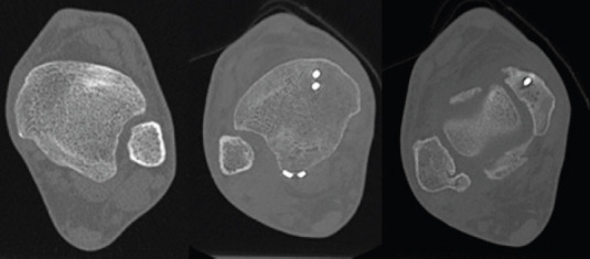
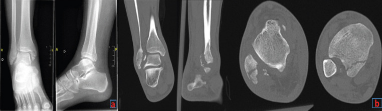
Case report: A 41-year-old male presented after a bicycle accident with a complex ankle fracture involving a transverse medial malleolus fracture, a postero-medial tibial fragment, a fibular tip fracture, and two additional posterior fibular fragments. Despite initial fracture management, including closed reduction and open fixation, the patient developed chronic pain and instability due to malreduction. Computed tomography imaging revealed instability of the fibula within the fibular notch, indicating syndesmotic instability. The surgical procedure included fibular osteotomy, temporary fixation with K-wires, syndesmotic fixation with the TightRope® system, and PITFL repair using the InternalBrace™ ligament augmentation system. Intraoperative three-dimensional imaging confirmed successful reduction and stabilization.
Conclusion: Fibular avulsion of the PITFL is rare. Failure to diagnose the lesion may lead to malreduction of the fibula within the incisura. The combination of osteotomy, TightRope® syndesmosis fixation, and InternalBrace™ PITFL repair provides a reliable option for managing complex PITFL injuries.


 求助内容:
求助内容: 应助结果提醒方式:


