{"title":"Disseminated Skeletal Cryptococcosis: A Case Report.","authors":"David Joseph, Prannoy Paul, Vishnu Padmanabhan, Athul Rajesh, Raj Vignesh, Raphael Baby","doi":"10.13107/jocr.2025.v15.i08.5912","DOIUrl":null,"url":null,"abstract":"<p><strong>Introduction: </strong>Cryptococcus is a rare cause of osteomyelitis, especially in immunocompromised individuals. This case report discusses a rare case of disseminated cryptococcosis with multiple bone lesions in a patient with isolated CD4 lymphocytopenia.</p><p><strong>Case report: </strong>A 31-year-old apparently normal Indian male presented with pain and swelling of his right proximal femur for 8 months without any history of trauma. He also reported a similar swelling in his chest wall with allergic respiratory symptoms for 8 years. Laboratory analysis revealed mild elevation in inflammatory markers. Magnetic resonance imaging of the pelvis revealed osteolytic lesions in the right proximal femur and pubic bone with soft tissue collections, and computed tomography scan of the chest showed an osteolytic lesion in the right 9th rib with an overlying soft tissue collection and a subpleural cavitary nodule in the left lower lobe posterior basal segment. Although initially treated as a case of clinically diagnosed tuberculosis, the patient did not get any relief with antitubercular therapy. Fine needle aspiration cytology and fungal culture identified Cryptococcus neoformans from both lesions and from the blood culture. The patient responded well to antifungal treatment and is currently symptom free.</p><p><strong>Conclusion: </strong>Cryptococcosis should be considered as a rare differential diagnosis in patients presenting with bone pain and multiple lytic lesions. Definitive diagnosis requires a fungal culture from the affected areas. Early treatment with antifungals is important in preventing complications and death.</p>","PeriodicalId":16647,"journal":{"name":"Journal of Orthopaedic Case Reports","volume":"15 8","pages":"130-134"},"PeriodicalIF":0.0000,"publicationDate":"2025-08-01","publicationTypes":"Journal Article","fieldsOfStudy":null,"isOpenAccess":false,"openAccessPdf":"https://www.ncbi.nlm.nih.gov/pmc/articles/PMC12328977/pdf/","citationCount":"0","resultStr":null,"platform":"Semanticscholar","paperid":null,"PeriodicalName":"Journal of Orthopaedic Case Reports","FirstCategoryId":"1085","ListUrlMain":"https://doi.org/10.13107/jocr.2025.v15.i08.5912","RegionNum":0,"RegionCategory":null,"ArticlePicture":[],"TitleCN":null,"AbstractTextCN":null,"PMCID":null,"EPubDate":"","PubModel":"","JCR":"","JCRName":"","Score":null,"Total":0}
引用次数: 0
Abstract
Introduction: Cryptococcus is a rare cause of osteomyelitis, especially in immunocompromised individuals. This case report discusses a rare case of disseminated cryptococcosis with multiple bone lesions in a patient with isolated CD4 lymphocytopenia.
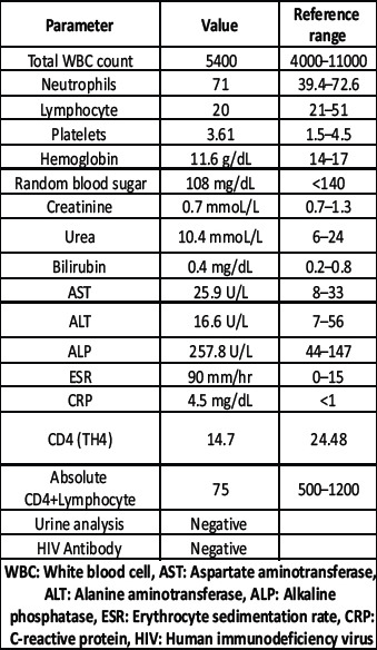
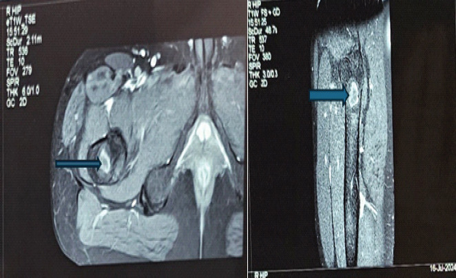
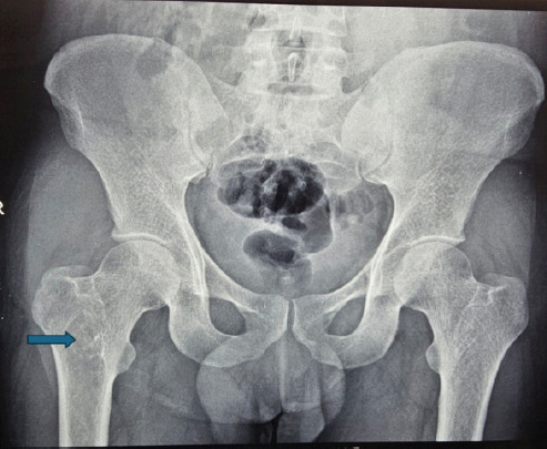
Case report: A 31-year-old apparently normal Indian male presented with pain and swelling of his right proximal femur for 8 months without any history of trauma. He also reported a similar swelling in his chest wall with allergic respiratory symptoms for 8 years. Laboratory analysis revealed mild elevation in inflammatory markers. Magnetic resonance imaging of the pelvis revealed osteolytic lesions in the right proximal femur and pubic bone with soft tissue collections, and computed tomography scan of the chest showed an osteolytic lesion in the right 9th rib with an overlying soft tissue collection and a subpleural cavitary nodule in the left lower lobe posterior basal segment. Although initially treated as a case of clinically diagnosed tuberculosis, the patient did not get any relief with antitubercular therapy. Fine needle aspiration cytology and fungal culture identified Cryptococcus neoformans from both lesions and from the blood culture. The patient responded well to antifungal treatment and is currently symptom free.
Conclusion: Cryptococcosis should be considered as a rare differential diagnosis in patients presenting with bone pain and multiple lytic lesions. Definitive diagnosis requires a fungal culture from the affected areas. Early treatment with antifungals is important in preventing complications and death.

 求助内容:
求助内容: 应助结果提醒方式:


