Comparative analysis of superficial collecting lymph vessels in normal tissue and lymphedema using video-capillaroscopy
IF 2.7
4区 医学
Q2 PERIPHERAL VASCULAR DISEASE
引用次数: 0
Abstract
Introduction
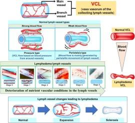
Lymphedema is a chronic, progressive disorder characterized by impaired lymphatic transport, tissue swelling, and fibrosis. This study used video-capillaroscopy, a high-resolution imaging technique, to assess superficial collecting lymphatic vessels and their vasa vasorum in patients with lymphedema. By comparing these microvascular structures to those in healthy tissue, we aimed to identify early vascular changes contributing to disease progression.
Methods
VC recordings were retrospectively analyzed from 28 limbs in 17 patients with lower extremity lymphedema and 53 lymphatic vessels in 12 cancer patients undergoing reconstructive surgery. In the latter group, observations were performed on normal subcutaneous tissue exposed at the donor site during flap harvest. These areas showed no tumor involvement, regional metastasis, or prior chemotherapy/radiotherapy. Thus, all normal tissue observations were made on untreated, unaffected sites. VCLs were classified into six stages (0–5) based on morphology and flow. Vessel diameter and red blood cell (RBC) velocity were measured. Statistical significance was set at p < 0.05.
Results
In normal donor tissue, mean VCL main vessel diameter and RBC velocity were 0.038 ± 0.031 mm and 185 ± 160.5 μm/s. In lymphedema, these values were reduced to 0.033 ± 0.024 mm and 28.3 ± 36.8 μm/s (p < 0.001). VCL Stage 0 showed preserved flow (p = 0.178), while Stages 1–5 exhibited progressive impairment.
Conclusion
These findings suggest that early ischemic changes in the vasa vasorum may precede lymphatic dysfunction and fibrosis in lymphedema. Preserving microvascular integrity should be a therapeutic focus, alongside drainage support. Further studies are needed to clarify clinical relevance and optimize treatment strategies.
视频毛细血管镜下正常组织浅表集淋巴管与淋巴水肿的对比分析。
简介:淋巴水肿是一种慢性进行性疾病,以淋巴运输受损、组织肿胀和纤维化为特征。本研究使用视频毛细血管镜,一种高分辨率成像技术,来评估淋巴水肿患者的浅表收集淋巴管及其血管。通过将这些微血管结构与健康组织的微血管结构进行比较,我们旨在确定有助于疾病进展的早期血管变化。方法:回顾性分析17例下肢淋巴水肿患者28条肢体和12例肿瘤重建手术患者53条淋巴管的VC记录。在后一组中,观察在皮瓣收获期间暴露在供区正常皮下组织。这些区域未见肿瘤累及、局部转移或既往化疗/放疗。因此,所有正常组织的观察都是在未经治疗、未受影响的部位进行的。根据形态和流量将vcl分为6级(0-5级)。测量血管直径和红细胞流速。结果:在正常供体组织中,VCL主干血管直径和红细胞流速分别为0.038 ± 0.031 mm和185 ± 160.5 μm/s。在淋巴水肿中,这一数值分别降至0.033 ± 0.024 mm和28.3 ± 36.8 μm/s (p )。结论:这些结果提示,在淋巴水肿中,血管血管早期缺血改变可能早于淋巴功能障碍和纤维化。保持微血管的完整性应该是治疗的重点,同时支持引流。需要进一步的研究来阐明临床相关性和优化治疗策略。
本文章由计算机程序翻译,如有差异,请以英文原文为准。
求助全文
约1分钟内获得全文
求助全文
来源期刊
Microvascular research
医学-外周血管病
CiteScore
6.00
自引率
3.20%
发文量
158
审稿时长
43 days
期刊介绍:
Microvascular Research is dedicated to the dissemination of fundamental information related to the microvascular field. Full-length articles presenting the results of original research and brief communications are featured.
Research Areas include:
• Angiogenesis
• Biochemistry
• Bioengineering
• Biomathematics
• Biophysics
• Cancer
• Circulatory homeostasis
• Comparative physiology
• Drug delivery
• Neuropharmacology
• Microvascular pathology
• Rheology
• Tissue Engineering.
 求助内容:
求助内容: 应助结果提醒方式:


