Sepehr Aghajanian, Fateme Mohammadifard, Ida Mohammadi, Shahryar Rajai Firouzabadi, Ali Baradaran Bagheri, Elham Moases Ghaffary, Omid Mirmosayyeb
{"title":"Longitudinal structural MRI-based deep learning and radiomics features for predicting Alzheimer's disease progression.","authors":"Sepehr Aghajanian, Fateme Mohammadifard, Ida Mohammadi, Shahryar Rajai Firouzabadi, Ali Baradaran Bagheri, Elham Moases Ghaffary, Omid Mirmosayyeb","doi":"10.1186/s13195-025-01827-2","DOIUrl":null,"url":null,"abstract":"<p><strong>Background: </strong>Alzheimer's disease (AD) is the principal cause of dementia and requires the early diagnosis of people with mild cognitive impairment (MCI) who are at high risk of progressing. Early diagnosis is imperative for optimizing clinical management and selecting proper therapeutic interventions. Structural magnetic resonance imaging (MRI) markers have been widely investigated for predicting the conversion of MCI to AD, and recent advances in deep learning (DL) methods offer enhanced capabilities for identifying subtle neurodegenerative changes over time.</p><p><strong>Methods: </strong>We selected 228 MCI participants from the Alzheimer's Disease Neuroimaging Initiative (ADNI) who had at least three T1-weighted MRI scans within 18 months of baseline. MRI volumes underwent bias correction, segmentation, and radiomics feature extraction. A 3D residual network (ResNet3D) was trained using a pairwise ranking loss to capture single-timepoint risk scores. Longitudinal analyses were performed by extracting deep convolutional neural network (CNN) embeddings and gray matter radiomics for each scan, which were put into a time-aware long short-term memory (LSTM) model with an attention mechanism.</p><p><strong>Results: </strong>A single-timepoint ResNet3D model achieved modest performance (c-index ~ 0.70). Incorporating longitudinal MRI files or downstream survival models led to a pronounced prognostic improvement (c-index:0.80-0.90), but was not further improved by longitudinal radiomics data. Time-specific classification within two- and three-year and five-year windows after the last MRI acquisition showed high accuracy (AUC > 0.85). Several radiomics, including gray matter surface to volume and elongation, emerged as the most predictive features. Each SD change in the gray matter surface to volume change within the last visit was associated with an increased risk of developing AD (HR: 1.50; 95% CI: 1.25-1.79).</p><p><strong>Conclusions: </strong>These findings emphasize the value of structural MRI within the advanced DL architectures for predicting MCI-to-AD conversion. The approach may enable earlier risk stratification and targeted interventions for individuals most likely to progress. limitations in sample size and computational resources warrant larger, more diverse studies to confirm these observations and explore additional improvements.</p>","PeriodicalId":7516,"journal":{"name":"Alzheimer's Research & Therapy","volume":"17 1","pages":"182"},"PeriodicalIF":7.6000,"publicationDate":"2025-08-07","publicationTypes":"Journal Article","fieldsOfStudy":null,"isOpenAccess":false,"openAccessPdf":"https://www.ncbi.nlm.nih.gov/pmc/articles/PMC12330009/pdf/","citationCount":"0","resultStr":null,"platform":"Semanticscholar","paperid":null,"PeriodicalName":"Alzheimer's Research & Therapy","FirstCategoryId":"3","ListUrlMain":"https://doi.org/10.1186/s13195-025-01827-2","RegionNum":1,"RegionCategory":"医学","ArticlePicture":[],"TitleCN":null,"AbstractTextCN":null,"PMCID":null,"EPubDate":"","PubModel":"","JCR":"Q1","JCRName":"CLINICAL NEUROLOGY","Score":null,"Total":0}
引用次数: 0
Abstract
Background: Alzheimer's disease (AD) is the principal cause of dementia and requires the early diagnosis of people with mild cognitive impairment (MCI) who are at high risk of progressing. Early diagnosis is imperative for optimizing clinical management and selecting proper therapeutic interventions. Structural magnetic resonance imaging (MRI) markers have been widely investigated for predicting the conversion of MCI to AD, and recent advances in deep learning (DL) methods offer enhanced capabilities for identifying subtle neurodegenerative changes over time.
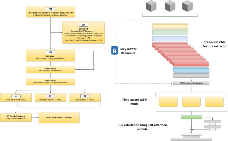
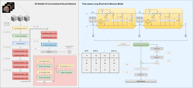
Methods: We selected 228 MCI participants from the Alzheimer's Disease Neuroimaging Initiative (ADNI) who had at least three T1-weighted MRI scans within 18 months of baseline. MRI volumes underwent bias correction, segmentation, and radiomics feature extraction. A 3D residual network (ResNet3D) was trained using a pairwise ranking loss to capture single-timepoint risk scores. Longitudinal analyses were performed by extracting deep convolutional neural network (CNN) embeddings and gray matter radiomics for each scan, which were put into a time-aware long short-term memory (LSTM) model with an attention mechanism.
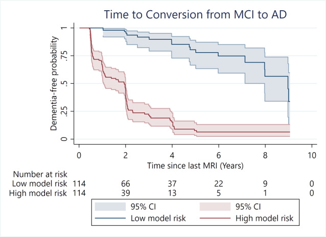
Results: A single-timepoint ResNet3D model achieved modest performance (c-index ~ 0.70). Incorporating longitudinal MRI files or downstream survival models led to a pronounced prognostic improvement (c-index:0.80-0.90), but was not further improved by longitudinal radiomics data. Time-specific classification within two- and three-year and five-year windows after the last MRI acquisition showed high accuracy (AUC > 0.85). Several radiomics, including gray matter surface to volume and elongation, emerged as the most predictive features. Each SD change in the gray matter surface to volume change within the last visit was associated with an increased risk of developing AD (HR: 1.50; 95% CI: 1.25-1.79).
Conclusions: These findings emphasize the value of structural MRI within the advanced DL architectures for predicting MCI-to-AD conversion. The approach may enable earlier risk stratification and targeted interventions for individuals most likely to progress. limitations in sample size and computational resources warrant larger, more diverse studies to confirm these observations and explore additional improvements.
期刊介绍:
Alzheimer's Research & Therapy is an international peer-reviewed journal that focuses on translational research into Alzheimer's disease and other neurodegenerative diseases. It publishes open-access basic research, clinical trials, drug discovery and development studies, and epidemiologic studies. The journal also includes reviews, viewpoints, commentaries, debates, and reports. All articles published in Alzheimer's Research & Therapy are included in several reputable databases such as CAS, Current contents, DOAJ, Embase, Journal Citation Reports/Science Edition, MEDLINE, PubMed, PubMed Central, Science Citation Index Expanded (Web of Science) and Scopus.


 求助内容:
求助内容: 应助结果提醒方式:


