Amreen Shakur, Grant D Stewart, Timothy J Sadler, Judith L Babar, Anne Y Warren, Stephen Scullion, Abhishekh H Ashok, Sumit Karia, Igor Chipurovski, James Whitworth, Stefan J Marciniak, Eamonn R Maher, Maria Ta Wetscherek
{"title":"Pulmonary cysts as a diagnostic indicator of Birt-Hogg-Dubé syndrome in patients with renal neoplasm.","authors":"Amreen Shakur, Grant D Stewart, Timothy J Sadler, Judith L Babar, Anne Y Warren, Stephen Scullion, Abhishekh H Ashok, Sumit Karia, Igor Chipurovski, James Whitworth, Stefan J Marciniak, Eamonn R Maher, Maria Ta Wetscherek","doi":"10.1186/s13244-025-02053-y","DOIUrl":null,"url":null,"abstract":"<p><strong>Objectives: </strong>To assess the presence and CT features of pulmonary cysts (PCs) in patients with renal neoplasms (RN) as a hallmark of Birt-Hogg-Dubé syndrome (BHDS).</p><p><strong>Materials and methods: </strong>Single institution retrospective study of all patients with histological RN between May 2014 and May 2020. Individuals with non-renal neoplasm, nephroblastoma, benign cysts, < 18 years old, or without thoracic CT were excluded. Demographics, history of smoking, pneumothorax and cutaneous fibrofolliculomas/trichodischomas, family history of pneumothorax or RN, and genetic testing were recorded. Number, location, distribution and morphology of PCs were assessed on thoracic CT. Differences between patients with positive (BHD+) and negative (BHD-) genetics were analysed. An independent cohort of 10 BHDS patients was added to calculate the diagnostic accuracy of cyst features.</p><p><strong>Results: </strong>Of 1475 patients with RN, 127 (8.6%) had PCs; 40 underwent genetic testing (median age 56 [49-68], 28 men), and 6/127 (4.7%) individuals tested positive for BHDS. BHD+ had significantly more and larger cysts, affecting more lobes (p < 0.01). Higher prevalence of PCs with a perivascular (100% vs. 37%; p = 0.01) and interlobular septal location (100% vs. 16%; p < 0.001), and perilymphatic distribution (100% vs. 5%; p < 0.001) was found in BHD+. All BHD+ had elliptical, irregular, and variable shape PCs, compared to a lower prevalence of these in BHD- (p < 0.01). Traversing vein sign was more common in BHD+ (83% vs. 24%; p = 0.01). The highest accuracy was achieved for perilymphatic distribution (97%), followed by irregular shape (94%) and interlobular septal location (91%).</p><p><strong>Conclusion: </strong>Specific CT features of PC in patients with RN can be highly indicative of BHDS.</p><p><strong>Critical relevance statement: </strong>Radiologists can play a crucial role in the diagnosis of Birt-Hogg-Dubé syndrome (BHDS) by recognising specific CT features of pulmonary cysts; a diagnosis of BHDS has implications for family testing and timely, life-long screening for renal neoplasm.</p><p><strong>Key points: </strong>Birt-Hogg-Dubé syndrome (BHDS) should be considered in patients with renal neoplasms and multiple pulmonary cysts. A lower zone predominant, perilymphatic distribution of pulmonary cysts is a strong indicator of BHDS. Identifying specific CT features of pulmonary cysts can improve recognition of BHDS.</p>","PeriodicalId":13639,"journal":{"name":"Insights into Imaging","volume":"16 1","pages":"169"},"PeriodicalIF":4.5000,"publicationDate":"2025-08-06","publicationTypes":"Journal Article","fieldsOfStudy":null,"isOpenAccess":false,"openAccessPdf":"https://www.ncbi.nlm.nih.gov/pmc/articles/PMC12328879/pdf/","citationCount":"0","resultStr":null,"platform":"Semanticscholar","paperid":null,"PeriodicalName":"Insights into Imaging","FirstCategoryId":"3","ListUrlMain":"https://doi.org/10.1186/s13244-025-02053-y","RegionNum":2,"RegionCategory":"医学","ArticlePicture":[],"TitleCN":null,"AbstractTextCN":null,"PMCID":null,"EPubDate":"","PubModel":"","JCR":"Q1","JCRName":"RADIOLOGY, NUCLEAR MEDICINE & MEDICAL IMAGING","Score":null,"Total":0}
引用次数: 0
Abstract
Objectives: To assess the presence and CT features of pulmonary cysts (PCs) in patients with renal neoplasms (RN) as a hallmark of Birt-Hogg-Dubé syndrome (BHDS).
Materials and methods: Single institution retrospective study of all patients with histological RN between May 2014 and May 2020. Individuals with non-renal neoplasm, nephroblastoma, benign cysts, < 18 years old, or without thoracic CT were excluded. Demographics, history of smoking, pneumothorax and cutaneous fibrofolliculomas/trichodischomas, family history of pneumothorax or RN, and genetic testing were recorded. Number, location, distribution and morphology of PCs were assessed on thoracic CT. Differences between patients with positive (BHD+) and negative (BHD-) genetics were analysed. An independent cohort of 10 BHDS patients was added to calculate the diagnostic accuracy of cyst features.
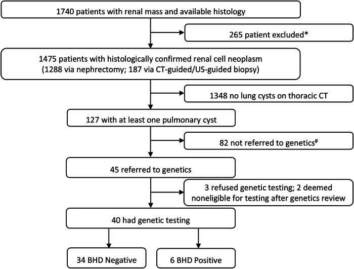
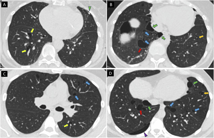
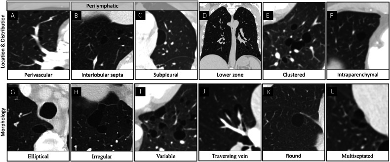
Results: Of 1475 patients with RN, 127 (8.6%) had PCs; 40 underwent genetic testing (median age 56 [49-68], 28 men), and 6/127 (4.7%) individuals tested positive for BHDS. BHD+ had significantly more and larger cysts, affecting more lobes (p < 0.01). Higher prevalence of PCs with a perivascular (100% vs. 37%; p = 0.01) and interlobular septal location (100% vs. 16%; p < 0.001), and perilymphatic distribution (100% vs. 5%; p < 0.001) was found in BHD+. All BHD+ had elliptical, irregular, and variable shape PCs, compared to a lower prevalence of these in BHD- (p < 0.01). Traversing vein sign was more common in BHD+ (83% vs. 24%; p = 0.01). The highest accuracy was achieved for perilymphatic distribution (97%), followed by irregular shape (94%) and interlobular septal location (91%).
Conclusion: Specific CT features of PC in patients with RN can be highly indicative of BHDS.
Critical relevance statement: Radiologists can play a crucial role in the diagnosis of Birt-Hogg-Dubé syndrome (BHDS) by recognising specific CT features of pulmonary cysts; a diagnosis of BHDS has implications for family testing and timely, life-long screening for renal neoplasm.
Key points: Birt-Hogg-Dubé syndrome (BHDS) should be considered in patients with renal neoplasms and multiple pulmonary cysts. A lower zone predominant, perilymphatic distribution of pulmonary cysts is a strong indicator of BHDS. Identifying specific CT features of pulmonary cysts can improve recognition of BHDS.
期刊介绍:
Insights into Imaging (I³) is a peer-reviewed open access journal published under the brand SpringerOpen. All content published in the journal is freely available online to anyone, anywhere!
I³ continuously updates scientific knowledge and progress in best-practice standards in radiology through the publication of original articles and state-of-the-art reviews and opinions, along with recommendations and statements from the leading radiological societies in Europe.
Founded by the European Society of Radiology (ESR), I³ creates a platform for educational material, guidelines and recommendations, and a forum for topics of controversy.
A balanced combination of review articles, original papers, short communications from European radiological congresses and information on society matters makes I³ an indispensable source for current information in this field.
I³ is owned by the ESR, however authors retain copyright to their article according to the Creative Commons Attribution License (see Copyright and License Agreement). All articles can be read, redistributed and reused for free, as long as the author of the original work is cited properly.
The open access fees (article-processing charges) for this journal are kindly sponsored by ESR for all Members.
The journal went open access in 2012, which means that all articles published since then are freely available online.


 求助内容:
求助内容: 应助结果提醒方式:


