{"title":"A convenient model based on mammography and magnetic resonance imaging for preoperative differentiation of breast phyllodes tumors and fibroadenomas.","authors":"Xiaowen Ma, Jinhui Li, Feixiang Hu, Yan Huang, Qin Xiao, Weijun Peng, Yajia Gu","doi":"10.21037/gs-2025-145","DOIUrl":null,"url":null,"abstract":"<p><strong>Background: </strong>Differentiation between breast phyllodes tumors (PTs) and fibroadenomas (FAs) remains a key clinical challenge, which is critical for formulating clinical treatment strategies. This study aimed to establish a fusion model based on mammography (MG) and magnetic resonance imaging (MRI) for the preoperative differentiation of PTs and FAs.</p><p><strong>Methods: </strong>The clinical data, MG images, and magnetic resonance (MR) images of patients with breast FAs treated in Fudan University Shanghai Cancer Center from October 2019 to December 2020, as well as patients with PTs treated from January 2011 to December 2020, were retrospectively collected. Univariate and multivariate logistic regression analyses were conducted to select independent factors and to construct a diagnostic model to differentiate PTs and FAs. The diagnostic performance of the model was evaluated using the receiver operating characteristic (ROC) curve, calibration curve, and decision curve analysis (DCA).</p><p><strong>Results: </strong>A total of 147 patients with FAs and 138 patients with PTs were included in this study. Patient age, maximum diameter of mass, density on MG images, lobulation on MR images, and time-intensity curve (TIC) were independent factors contributing to the differential diagnosis. Finally, the fusion model showed satisfactory discrimination [area under the curve (AUC) 0.90, 95% confidence interval (CI): 0.86-0.94] and calibration. DCA indicated good clinical benefit, as indicated by most values being within threshold probabilities.</p><p><strong>Conclusions: </strong>Breast MG and MRI findings help differentiate between FAs and PTs preoperatively. The multimodal fusion model was clinically efficacious and thus useful for accurate clinical diagnosis and treatment.</p>","PeriodicalId":12760,"journal":{"name":"Gland surgery","volume":"14 7","pages":"1306-1317"},"PeriodicalIF":1.6000,"publicationDate":"2025-07-31","publicationTypes":"Journal Article","fieldsOfStudy":null,"isOpenAccess":false,"openAccessPdf":"https://www.ncbi.nlm.nih.gov/pmc/articles/PMC12322753/pdf/","citationCount":"0","resultStr":null,"platform":"Semanticscholar","paperid":null,"PeriodicalName":"Gland surgery","FirstCategoryId":"3","ListUrlMain":"https://doi.org/10.21037/gs-2025-145","RegionNum":3,"RegionCategory":"医学","ArticlePicture":[],"TitleCN":null,"AbstractTextCN":null,"PMCID":null,"EPubDate":"2025/7/28 0:00:00","PubModel":"Epub","JCR":"Q3","JCRName":"SURGERY","Score":null,"Total":0}
引用次数: 0
Abstract
Background: Differentiation between breast phyllodes tumors (PTs) and fibroadenomas (FAs) remains a key clinical challenge, which is critical for formulating clinical treatment strategies. This study aimed to establish a fusion model based on mammography (MG) and magnetic resonance imaging (MRI) for the preoperative differentiation of PTs and FAs.
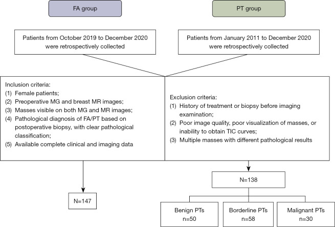
Methods: The clinical data, MG images, and magnetic resonance (MR) images of patients with breast FAs treated in Fudan University Shanghai Cancer Center from October 2019 to December 2020, as well as patients with PTs treated from January 2011 to December 2020, were retrospectively collected. Univariate and multivariate logistic regression analyses were conducted to select independent factors and to construct a diagnostic model to differentiate PTs and FAs. The diagnostic performance of the model was evaluated using the receiver operating characteristic (ROC) curve, calibration curve, and decision curve analysis (DCA).
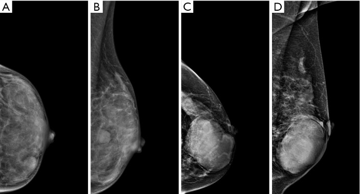
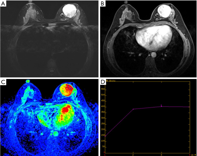
Results: A total of 147 patients with FAs and 138 patients with PTs were included in this study. Patient age, maximum diameter of mass, density on MG images, lobulation on MR images, and time-intensity curve (TIC) were independent factors contributing to the differential diagnosis. Finally, the fusion model showed satisfactory discrimination [area under the curve (AUC) 0.90, 95% confidence interval (CI): 0.86-0.94] and calibration. DCA indicated good clinical benefit, as indicated by most values being within threshold probabilities.
Conclusions: Breast MG and MRI findings help differentiate between FAs and PTs preoperatively. The multimodal fusion model was clinically efficacious and thus useful for accurate clinical diagnosis and treatment.
期刊介绍:
Gland Surgery (Gland Surg; GS, Print ISSN 2227-684X; Online ISSN 2227-8575) being indexed by PubMed/PubMed Central, is an open access, peer-review journal launched at May of 2012, published bio-monthly since February 2015.
 求助内容:
求助内容: 应助结果提醒方式:


