Ioana Hălmaciu, Anca Meda Văsieșiu, Andrei Manea, Andrei Dragomir, Ioana Tripon, Vlad Vunvulea, Cristian Boeriu, Andrea Rus, Minodora Dobreanu
{"title":"Artificial intelligence algorithms based approach in evaluating COVID-19 patients and management.","authors":"Ioana Hălmaciu, Anca Meda Văsieșiu, Andrei Manea, Andrei Dragomir, Ioana Tripon, Vlad Vunvulea, Cristian Boeriu, Andrea Rus, Minodora Dobreanu","doi":"10.2478/jccm-2025-0032","DOIUrl":null,"url":null,"abstract":"<p><strong>Introduction: </strong>COVID-19 pneumonia manifests with a wide range of clinical symptoms, from minor flu-like signs to multi-organ failure. Chest computed tomography (CT) is the most effective imaging method for assessing the extent of the pulmonary lesions and correlates with disease severity. Increased workloads during the COVID-19 pandemic led to the development of various artificial intelligence tools to enable quicker diagnoses and quantitative evaluations of the lesions.</p><p><strong>Aim of the study: </strong>This study aims to analyse the correlation between lung lesions identified on CT scans and the biological inflammatory markers assessed, to establish the survival rate among patients.</p><p><strong>Methods: </strong>This retrospective study included 120 patients diagnosed with moderate to severe COVID-19 pneumonia who were admitted to the intensive care unit and the internal medicine department between September 2020 and October 2021. Each patient underwent a chest CT scan, which was subsequently analysed by two radiologists and an AI post-processing software. On the same day, blood was collected from the patients to determine inflammatory markers. The markers analysed in this study include the neutrophil-lymphocyte ratio (NLR), monocyte-lymphocyte ratio, platelet-lymphocyte ratio, systemic immune-inflammatory index, systemic inflammation response index, systemic inflammation index, and serum interleukin-6 value.</p><p><strong>Results: </strong>There were strong and very strong correlations between the derived inflammatory markers, interleukin-6, and the CT severity scores obtained by the AI algorithm (r=0.851, p<0.001 in the case of NLR). Higher values of the inflammatory markers and high lung opacity scores correlated with a decreased survival rate. Crazy paving was also associated with an increased risk of mortality (OR=2.89, p=0.006).</p><p><strong>Conclusions: </strong>AI-based chest CT analysis plays a crucial role in assessing patients with COVID-19 pneumonia. When combined with inflammatory markers, it provides a reliable and objective method for evaluating COVID-19 pneumonia, enhancing the accuracy of diagnosis.</p>","PeriodicalId":44227,"journal":{"name":"Journal of Critical Care Medicine","volume":"11 3","pages":"247-256"},"PeriodicalIF":1.7000,"publicationDate":"2025-07-31","publicationTypes":"Journal Article","fieldsOfStudy":null,"isOpenAccess":false,"openAccessPdf":"https://www.ncbi.nlm.nih.gov/pmc/articles/PMC12321253/pdf/","citationCount":"0","resultStr":null,"platform":"Semanticscholar","paperid":null,"PeriodicalName":"Journal of Critical Care Medicine","FirstCategoryId":"1085","ListUrlMain":"https://doi.org/10.2478/jccm-2025-0032","RegionNum":0,"RegionCategory":null,"ArticlePicture":[],"TitleCN":null,"AbstractTextCN":null,"PMCID":null,"EPubDate":"2025/7/1 0:00:00","PubModel":"eCollection","JCR":"Q4","JCRName":"CRITICAL CARE MEDICINE","Score":null,"Total":0}
引用次数: 0
Abstract
Introduction: COVID-19 pneumonia manifests with a wide range of clinical symptoms, from minor flu-like signs to multi-organ failure. Chest computed tomography (CT) is the most effective imaging method for assessing the extent of the pulmonary lesions and correlates with disease severity. Increased workloads during the COVID-19 pandemic led to the development of various artificial intelligence tools to enable quicker diagnoses and quantitative evaluations of the lesions.
Aim of the study: This study aims to analyse the correlation between lung lesions identified on CT scans and the biological inflammatory markers assessed, to establish the survival rate among patients.
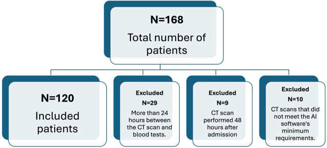
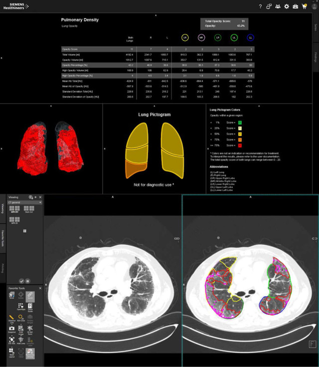
Methods: This retrospective study included 120 patients diagnosed with moderate to severe COVID-19 pneumonia who were admitted to the intensive care unit and the internal medicine department between September 2020 and October 2021. Each patient underwent a chest CT scan, which was subsequently analysed by two radiologists and an AI post-processing software. On the same day, blood was collected from the patients to determine inflammatory markers. The markers analysed in this study include the neutrophil-lymphocyte ratio (NLR), monocyte-lymphocyte ratio, platelet-lymphocyte ratio, systemic immune-inflammatory index, systemic inflammation response index, systemic inflammation index, and serum interleukin-6 value.
Results: There were strong and very strong correlations between the derived inflammatory markers, interleukin-6, and the CT severity scores obtained by the AI algorithm (r=0.851, p<0.001 in the case of NLR). Higher values of the inflammatory markers and high lung opacity scores correlated with a decreased survival rate. Crazy paving was also associated with an increased risk of mortality (OR=2.89, p=0.006).
Conclusions: AI-based chest CT analysis plays a crucial role in assessing patients with COVID-19 pneumonia. When combined with inflammatory markers, it provides a reliable and objective method for evaluating COVID-19 pneumonia, enhancing the accuracy of diagnosis.

 求助内容:
求助内容: 应助结果提醒方式:


