Tjeerd van der Veer, Eleni-Rosalina Andrinopoulou, Punitkumar Makani, Gert-Jan Braunstahl, Harm A W M Tiddens
{"title":"Automated computed tomographic analysis of bronchial thickness and mucus plugs in bronchiectasis with asthma.","authors":"Tjeerd van der Veer, Eleni-Rosalina Andrinopoulou, Punitkumar Makani, Gert-Jan Braunstahl, Harm A W M Tiddens","doi":"10.1183/23120541.00736-2024","DOIUrl":null,"url":null,"abstract":"<p><strong>Background: </strong>Bronchiectasis disease is characterised by cough, sputum and exacerbations, with chest computed tomography (CT) typically showing bronchial wall thickening and mucus plugging in addition to bronchial dilation. Asthma is a common comorbidity and associated with increased, eosinophilic, airway inflammation. Automated measurements of bronchial wall thickening and mucus plugs may serve as biomarkers for inflammation and are associated with clinical characteristics such as spirometry, blood eosinophil counts and disease severity in patients with bronchiectasis and asthma co-diagnosis.</p><p><strong>Methods: </strong>In a cross-sectional retrospective cohort of 64 patients with bronchiectasis disease and asthma, we applied automated image analysis to assess bronchial dimensions and mucus plug metrics on chest CT scans. These metrics were correlated with spirometry, blood eosinophil counts as well as FACED and Bronchiectasis Severity Index (BSI) scores using correlations and multiple regression analyses.</p><p><strong>Results: </strong>In 63 patients, bronchial wall thickness and mucus plugs were quantified. Negative correlations were observed between bronchial wall thickness markers and spirometry (bronchial wall thickness/accompanying artery diameter and forced expiratory volume in 1 s (FEV<sub>1</sub>), r= -0.37; FEV<sub>1</sub>/forced vital capacity, r= -0.30). Mucus plugs correlated negatively with spirometry and positively with FACED and BSI scores (number of mucus plugs and BSI, r=0.45). Correlations with blood eosinophil counts were very weak. In multiple regression analyses, independent associations were observed for FEV<sub>1</sub>, <i>Pseudomonas aeruginosa</i> and frequent exacerbations.</p><p><strong>Conclusion: </strong>This study identified key relationships between automated measurements of bronchial wall thickness and mucus plugs and clinical characteristics, highlighting their potential as imaging biomarkers to enhance phenotyping, improve risk assessment and facilitate tailored treatment strategies in bronchiectasis.</p>","PeriodicalId":11739,"journal":{"name":"ERJ Open Research","volume":"11 4","pages":""},"PeriodicalIF":4.0000,"publicationDate":"2025-08-04","publicationTypes":"Journal Article","fieldsOfStudy":null,"isOpenAccess":false,"openAccessPdf":"https://www.ncbi.nlm.nih.gov/pmc/articles/PMC12320113/pdf/","citationCount":"0","resultStr":null,"platform":"Semanticscholar","paperid":null,"PeriodicalName":"ERJ Open Research","FirstCategoryId":"3","ListUrlMain":"https://doi.org/10.1183/23120541.00736-2024","RegionNum":3,"RegionCategory":"医学","ArticlePicture":[],"TitleCN":null,"AbstractTextCN":null,"PMCID":null,"EPubDate":"2025/7/1 0:00:00","PubModel":"eCollection","JCR":"Q1","JCRName":"RESPIRATORY SYSTEM","Score":null,"Total":0}
引用次数: 0
Abstract
Background: Bronchiectasis disease is characterised by cough, sputum and exacerbations, with chest computed tomography (CT) typically showing bronchial wall thickening and mucus plugging in addition to bronchial dilation. Asthma is a common comorbidity and associated with increased, eosinophilic, airway inflammation. Automated measurements of bronchial wall thickening and mucus plugs may serve as biomarkers for inflammation and are associated with clinical characteristics such as spirometry, blood eosinophil counts and disease severity in patients with bronchiectasis and asthma co-diagnosis.
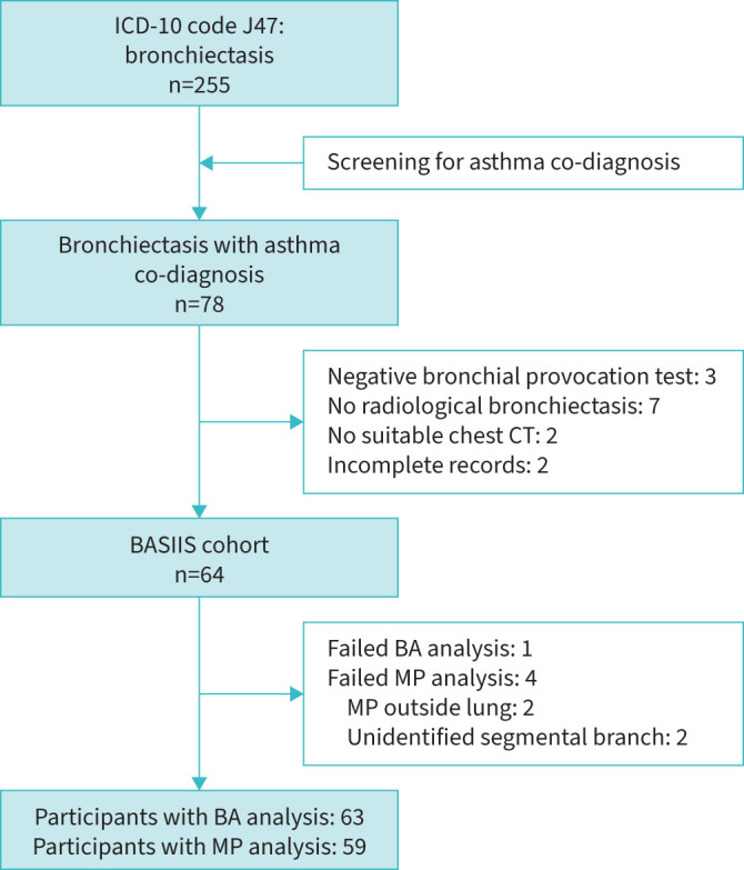
Methods: In a cross-sectional retrospective cohort of 64 patients with bronchiectasis disease and asthma, we applied automated image analysis to assess bronchial dimensions and mucus plug metrics on chest CT scans. These metrics were correlated with spirometry, blood eosinophil counts as well as FACED and Bronchiectasis Severity Index (BSI) scores using correlations and multiple regression analyses.
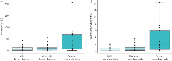
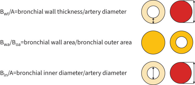
Results: In 63 patients, bronchial wall thickness and mucus plugs were quantified. Negative correlations were observed between bronchial wall thickness markers and spirometry (bronchial wall thickness/accompanying artery diameter and forced expiratory volume in 1 s (FEV1), r= -0.37; FEV1/forced vital capacity, r= -0.30). Mucus plugs correlated negatively with spirometry and positively with FACED and BSI scores (number of mucus plugs and BSI, r=0.45). Correlations with blood eosinophil counts were very weak. In multiple regression analyses, independent associations were observed for FEV1, Pseudomonas aeruginosa and frequent exacerbations.
Conclusion: This study identified key relationships between automated measurements of bronchial wall thickness and mucus plugs and clinical characteristics, highlighting their potential as imaging biomarkers to enhance phenotyping, improve risk assessment and facilitate tailored treatment strategies in bronchiectasis.
期刊介绍:
ERJ Open Research is a fully open access original research journal, published online by the European Respiratory Society. The journal aims to publish high-quality work in all fields of respiratory science and medicine, covering basic science, clinical translational science and clinical medicine. The journal was created to help fulfil the ERS objective to disseminate scientific and educational material to its members and to the medical community, but also to provide researchers with an affordable open access specialty journal in which to publish their work.

 求助内容:
求助内容: 应助结果提醒方式:


