Carlos D Pargas-Colina, Tori J Coble, Sara E Davis, David D Spence, Jonathan K Rowland, Derek M Kelly, Benjamin W Sheffer
{"title":"Outcomes of Suprapatellar Intramedullary Nail Fixation of Tibial Shaft Fractures in Skeletally Immature Patients.","authors":"Carlos D Pargas-Colina, Tori J Coble, Sara E Davis, David D Spence, Jonathan K Rowland, Derek M Kelly, Benjamin W Sheffer","doi":"10.1016/j.jposna.2025.100200","DOIUrl":null,"url":null,"abstract":"<p><strong>Background: </strong>Tibial shaft fractures in children require individualized treatment approaches, including elastic nails, plates and screws, intramedullary rigid nails (IMN), and external fixators, with selection based on fracture characteristics and patient factors. IMN risks damaging the physis, but can provide enhanced stability and immediate weight bearing. While both infrapatellar and suprapatellar approaches exist for IMN placement, studies in adults have demonstrated that the suprapatellar approach offers higher patient satisfaction, less anterior knee pain, and lower radiation exposure. However, there is a lack of data on IMN for pediatric tibial fractures.</p><p><strong>Methods: </strong>This single-center, retrospective cohort study analyzed patients aged 11-16 with open proximal tibia physis and tibial shaft fractures treated with IMN via the suprapatellar approach from January 2016 to October 2023. Demographic, fracture, and operative data were collected, and proximal tibial angles were measured. Follow-up radiographs were evaluated for tibial growth and alignment, with malunion defined as over 5 degrees of angular deformity.</p><p><strong>Results: </strong>Thirty-five patients aged 11 to 16 (mean 14.7 years, SD 1.1) met the inclusion criteria. Males comprised 60%; the average follow-up was 12.4 months. Most fractures (74%, 26/35) were closed. The most common (57%) fracture pattern was OA42A1-3. Fracture union occurred by 9 weeks in 79% of cases. The mechanical Medial Proximal Tibial Angle (mMPTA, range 85-90 degrees) and Posterior Proximal Tibial Angle (PPTA, range 77-84 degrees) were normal in all pre-operative patients; there were no statistically significant changes in mMPTA and PPTA over the follow-up period. The reported Visual Analog Scale pain scale averaged 1.87 at the final follow-up. Most patients reported return to full activities (87%), with the operative leg comparable to the contralateral leg in 78% of cases.</p><p><strong>Conclusions: </strong>Our findings demonstrated that suprapatellar IMN insertion leads to low pain scores and good function in short-term follow-up for adolescent patients treated for tibial shaft fractures. No proximal tibia growth disturbance was detected. Further prospective studies are warranted.</p><p><strong>Key concepts: </strong>(1)The suprapatellar approach appears to be safe for adolescent tibial nailing.(2)Growth disturbance was not encountered in this carefully selected patient group.(3)Radiographic tibial fracture union was achieved in call patients in this cohort.</p><p><strong>Level of evidence: </strong>Level IV.</p>","PeriodicalId":520850,"journal":{"name":"Journal of the Pediatric Orthopaedic Society of North America","volume":"12 ","pages":"100200"},"PeriodicalIF":0.0000,"publicationDate":"2025-05-23","publicationTypes":"Journal Article","fieldsOfStudy":null,"isOpenAccess":false,"openAccessPdf":"https://www.ncbi.nlm.nih.gov/pmc/articles/PMC12317414/pdf/","citationCount":"0","resultStr":null,"platform":"Semanticscholar","paperid":null,"PeriodicalName":"Journal of the Pediatric Orthopaedic Society of North America","FirstCategoryId":"1085","ListUrlMain":"https://doi.org/10.1016/j.jposna.2025.100200","RegionNum":0,"RegionCategory":null,"ArticlePicture":[],"TitleCN":null,"AbstractTextCN":null,"PMCID":null,"EPubDate":"2025/8/1 0:00:00","PubModel":"eCollection","JCR":"","JCRName":"","Score":null,"Total":0}
引用次数: 0
Abstract
Background: Tibial shaft fractures in children require individualized treatment approaches, including elastic nails, plates and screws, intramedullary rigid nails (IMN), and external fixators, with selection based on fracture characteristics and patient factors. IMN risks damaging the physis, but can provide enhanced stability and immediate weight bearing. While both infrapatellar and suprapatellar approaches exist for IMN placement, studies in adults have demonstrated that the suprapatellar approach offers higher patient satisfaction, less anterior knee pain, and lower radiation exposure. However, there is a lack of data on IMN for pediatric tibial fractures.
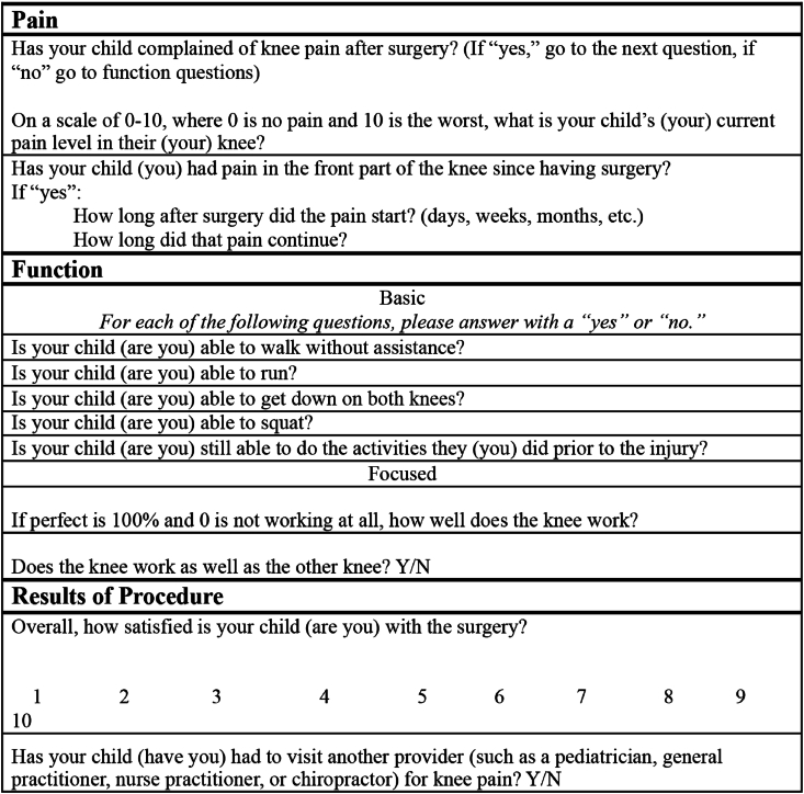
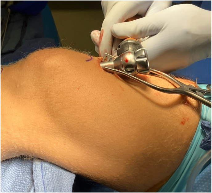
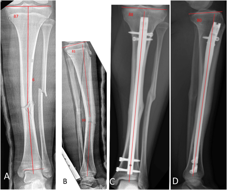
Methods: This single-center, retrospective cohort study analyzed patients aged 11-16 with open proximal tibia physis and tibial shaft fractures treated with IMN via the suprapatellar approach from January 2016 to October 2023. Demographic, fracture, and operative data were collected, and proximal tibial angles were measured. Follow-up radiographs were evaluated for tibial growth and alignment, with malunion defined as over 5 degrees of angular deformity.
Results: Thirty-five patients aged 11 to 16 (mean 14.7 years, SD 1.1) met the inclusion criteria. Males comprised 60%; the average follow-up was 12.4 months. Most fractures (74%, 26/35) were closed. The most common (57%) fracture pattern was OA42A1-3. Fracture union occurred by 9 weeks in 79% of cases. The mechanical Medial Proximal Tibial Angle (mMPTA, range 85-90 degrees) and Posterior Proximal Tibial Angle (PPTA, range 77-84 degrees) were normal in all pre-operative patients; there were no statistically significant changes in mMPTA and PPTA over the follow-up period. The reported Visual Analog Scale pain scale averaged 1.87 at the final follow-up. Most patients reported return to full activities (87%), with the operative leg comparable to the contralateral leg in 78% of cases.
Conclusions: Our findings demonstrated that suprapatellar IMN insertion leads to low pain scores and good function in short-term follow-up for adolescent patients treated for tibial shaft fractures. No proximal tibia growth disturbance was detected. Further prospective studies are warranted.
Key concepts: (1)The suprapatellar approach appears to be safe for adolescent tibial nailing.(2)Growth disturbance was not encountered in this carefully selected patient group.(3)Radiographic tibial fracture union was achieved in call patients in this cohort.

 求助内容:
求助内容: 应助结果提醒方式:


