Efficacy and safety of Bio 3D conduits composed of human umbilical cord-derived mesenchymal stromal cells: A proof-of-concept study in a canine ulnar nerve defect model.
{"title":"Efficacy and safety of Bio 3D conduits composed of human umbilical cord-derived mesenchymal stromal cells: A proof-of-concept study in a canine ulnar nerve defect model.","authors":"Kazuaki Fujita, Ryosuke Ikeguchi, Tomoki Aoyama, Takashi Noguchi, Koichi Yoshimoto, Daichi Sakamoto, Terunobu Iwai, Tetsuya Miyamoto, Yudai Miyazaki, Shizuka Akieda, Tokiko Nagamura-Inoue, Fumitaka Nagamura, Koichi Nakayama, Shuichi Matsuda","doi":"10.1177/09636897251361711","DOIUrl":null,"url":null,"abstract":"<p><p>Peripheral nerve injuries involving nerve defects remain challenging to treat. Although autologous nerve grafting is considered the gold standard, it has notable limitations, including donor site morbidity. To address this, we developed a scaffold-free Bio 3D conduit composed of human umbilical cord-derived mesenchymal stromal cells (UC-MSCs) using bioprinting technology. In this study, we evaluated its efficacy and safety in a canine ulnar nerve defect model. At 10 weeks postoperatively, the Bio 3D group showed better motor and sensory recovery compared with the allograft group, as demonstrated by the pinprick test, electrophysiological studies, and hypothenar muscle wet weight (0.978 ± 0.100 vs. 0.637 ± 0.151, n = 3). Morphometric analysis revealed greater axonal regeneration, including larger myelinated axon diameters (4.27 ± 0.342 µm vs. 3.69 ± 0.161 µm, n = 3) and thicker myelin sheaths (0.621 ± 0.088 µm vs. 0.497 ± 0.021 µm, n = 3). Immunostaining showed that the number of transplanted UC-MSCs diminished over time, likely after exerting their therapeutic effects. No adverse events, systemic abnormalities, or distant human cell migration was observed. These findings suggest that UC-MSC-derived Bio 3D conduits are a promising alternative for peripheral nerve regeneration, especially for patients wishing to avoid donor nerve harvesting.</p>","PeriodicalId":9721,"journal":{"name":"Cell Transplantation","volume":"34 ","pages":"9636897251361711"},"PeriodicalIF":3.2000,"publicationDate":"2025-01-01","publicationTypes":"Journal Article","fieldsOfStudy":null,"isOpenAccess":false,"openAccessPdf":"https://www.ncbi.nlm.nih.gov/pmc/articles/PMC12322347/pdf/","citationCount":"0","resultStr":null,"platform":"Semanticscholar","paperid":null,"PeriodicalName":"Cell Transplantation","FirstCategoryId":"3","ListUrlMain":"https://doi.org/10.1177/09636897251361711","RegionNum":4,"RegionCategory":"医学","ArticlePicture":[],"TitleCN":null,"AbstractTextCN":null,"PMCID":null,"EPubDate":"2025/8/3 0:00:00","PubModel":"Epub","JCR":"Q3","JCRName":"CELL & TISSUE ENGINEERING","Score":null,"Total":0}
引用次数: 0
Abstract
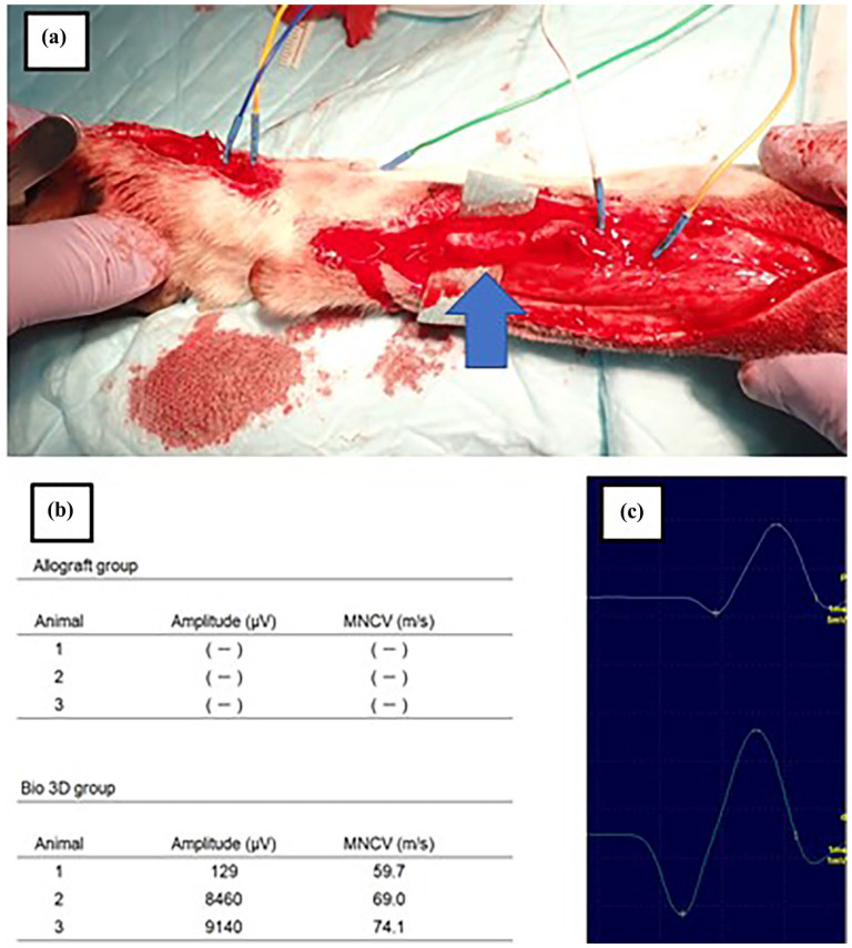
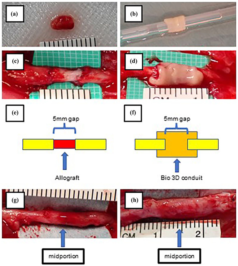
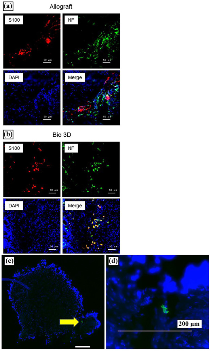
Peripheral nerve injuries involving nerve defects remain challenging to treat. Although autologous nerve grafting is considered the gold standard, it has notable limitations, including donor site morbidity. To address this, we developed a scaffold-free Bio 3D conduit composed of human umbilical cord-derived mesenchymal stromal cells (UC-MSCs) using bioprinting technology. In this study, we evaluated its efficacy and safety in a canine ulnar nerve defect model. At 10 weeks postoperatively, the Bio 3D group showed better motor and sensory recovery compared with the allograft group, as demonstrated by the pinprick test, electrophysiological studies, and hypothenar muscle wet weight (0.978 ± 0.100 vs. 0.637 ± 0.151, n = 3). Morphometric analysis revealed greater axonal regeneration, including larger myelinated axon diameters (4.27 ± 0.342 µm vs. 3.69 ± 0.161 µm, n = 3) and thicker myelin sheaths (0.621 ± 0.088 µm vs. 0.497 ± 0.021 µm, n = 3). Immunostaining showed that the number of transplanted UC-MSCs diminished over time, likely after exerting their therapeutic effects. No adverse events, systemic abnormalities, or distant human cell migration was observed. These findings suggest that UC-MSC-derived Bio 3D conduits are a promising alternative for peripheral nerve regeneration, especially for patients wishing to avoid donor nerve harvesting.
周围神经损伤包括神经缺损的治疗仍然具有挑战性。虽然自体神经移植被认为是金标准,但它有明显的局限性,包括供体部位的发病率。为了解决这个问题,我们利用生物打印技术开发了一种由人脐带来源的间充质基质细胞(UC-MSCs)组成的无支架生物3D导管。在本研究中,我们评估了其在犬尺神经缺损模型中的有效性和安全性。术后10周,针刺试验、电生理研究和鱼际下肌肉湿重(0.978±0.100 vs 0.637±0.151,n = 3)显示,Bio 3D组的运动和感觉恢复优于同种异体移植物组。形态计量学分析显示轴突再生更大,包括髓鞘轴突直径更大(4.27±0.342µm vs. 3.69±0.161µm, n = 3)和髓鞘更厚(0.621±0.088µm vs. 0.497±0.021µm, n = 3)。免疫染色显示移植的UC-MSCs数量随着时间的推移而减少,可能是在发挥其治疗作用后。没有观察到不良事件、全身异常或远处的人类细胞迁移。这些发现表明,uc - msc衍生的生物3D导管是周围神经再生的一种有希望的替代方法,特别是对于希望避免供体神经采集的患者。
期刊介绍:
Cell Transplantation, The Regenerative Medicine Journal is an open access, peer reviewed journal that is published 12 times annually. Cell Transplantation is a multi-disciplinary forum for publication of articles on cell transplantation and its applications to human diseases. Articles focus on a myriad of topics including the physiological, medical, pre-clinical, tissue engineering, stem cell, and device-oriented aspects of the nervous, endocrine, cardiovascular, and endothelial systems, as well as genetically engineered cells. Cell Transplantation also reports on relevant technological advances, clinical studies, and regulatory considerations related to the implantation of cells into the body in order to provide complete coverage of the field.
 求助内容:
求助内容: 应助结果提醒方式:


