{"title":"Deep learning-based super-resolution US radiomics to differentiate testicular seminoma and non-seminoma: an international multicenter study.","authors":"Yafang Zhang, Shilin Lu, Chuan Peng, Shichong Zhou, Irene Campo, Michele Bertolotto, Qian Li, Zhiyuan Wang, Dong Xu, Yun Wang, Jinshun Xu, Qinfu Wu, Xiaoying Hu, Wei Zheng, Jianhua Zhou","doi":"10.1186/s13244-025-02045-y","DOIUrl":null,"url":null,"abstract":"<p><strong>Objectives: </strong>Subvariants of testicular germ cell tumor (TGCT) significantly affect therapeutic strategies and patient prognosis. However, preoperatively distinguishing seminoma (SE) from non-seminoma (n-SE) remains a challenge. This study aimed to evaluate the performance of a deep learning-based super-resolution (SR) US radiomics model for SE/n-SE differentiation.</p><p><strong>Materials and methods: </strong>This international multicenter retrospective study recruited patients with confirmed TGCT between 2015 and 2023. A pre-trained SR reconstruction algorithm was applied to enhance native resolution (NR) images. NR and SR radiomics models were constructed, and the superior model was then integrated with clinical features to construct clinical-radiomics models. Diagnostic performance was evaluated by ROC analysis (AUC) and compared with radiologists' assessments using the DeLong test.</p><p><strong>Results: </strong>A total of 486 male patients were enrolled for training (n = 338), domestic (n = 92), and international (n = 59) validation sets. The SR radiomics model achieved AUCs of 0.90, 0.82, and 0.91, respectively, in the training, domestic, and international validation sets, significantly surpassing the NR model (p < 0.001, p = 0.031, and p = 0.001, respectively). The clinical-radiomics model exhibited a significantly higher across both domestic and international validation sets compared to the SR radiomics model alone (0.95 vs 0.82, p = 0.004; 0.97 vs 0.91, p = 0.031). Moreover, the clinical-radiomics model surpassed the performance of experienced radiologists in both domestic (AUC, 0.95 vs 0.85, p = 0.012) and international (AUC, 0.97 vs 0.77, p < 0.001) validation cohorts.</p><p><strong>Conclusions: </strong>The SR-based clinical-radiomics model can effectively differentiate between SE and n-SE.</p><p><strong>Critical relevance statement: </strong>This international multicenter study demonstrated that a radiomics model of deep learning-based SR reconstructed US images enabled effective differentiation between SE and n-SE.</p><p><strong>Key points: </strong>Clinical parameters and radiologists' assessments exhibit limited diagnostic accuracy for SE/n-SE differentiation in TGCT. Based on scrotal US images of TGCT, the SR radiomics models performed better than the NR radiomics models. The SR-based clinical-radiomics model outperforms both the radiomics model and radiologists' assessment, enabling accurate, non-invasive preoperative differentiation between SE and n-SE.</p>","PeriodicalId":13639,"journal":{"name":"Insights into Imaging","volume":"16 1","pages":"165"},"PeriodicalIF":4.5000,"publicationDate":"2025-08-01","publicationTypes":"Journal Article","fieldsOfStudy":null,"isOpenAccess":false,"openAccessPdf":"https://www.ncbi.nlm.nih.gov/pmc/articles/PMC12316629/pdf/","citationCount":"0","resultStr":null,"platform":"Semanticscholar","paperid":null,"PeriodicalName":"Insights into Imaging","FirstCategoryId":"3","ListUrlMain":"https://doi.org/10.1186/s13244-025-02045-y","RegionNum":2,"RegionCategory":"医学","ArticlePicture":[],"TitleCN":null,"AbstractTextCN":null,"PMCID":null,"EPubDate":"","PubModel":"","JCR":"Q1","JCRName":"RADIOLOGY, NUCLEAR MEDICINE & MEDICAL IMAGING","Score":null,"Total":0}
引用次数: 0
Abstract
Objectives: Subvariants of testicular germ cell tumor (TGCT) significantly affect therapeutic strategies and patient prognosis. However, preoperatively distinguishing seminoma (SE) from non-seminoma (n-SE) remains a challenge. This study aimed to evaluate the performance of a deep learning-based super-resolution (SR) US radiomics model for SE/n-SE differentiation.
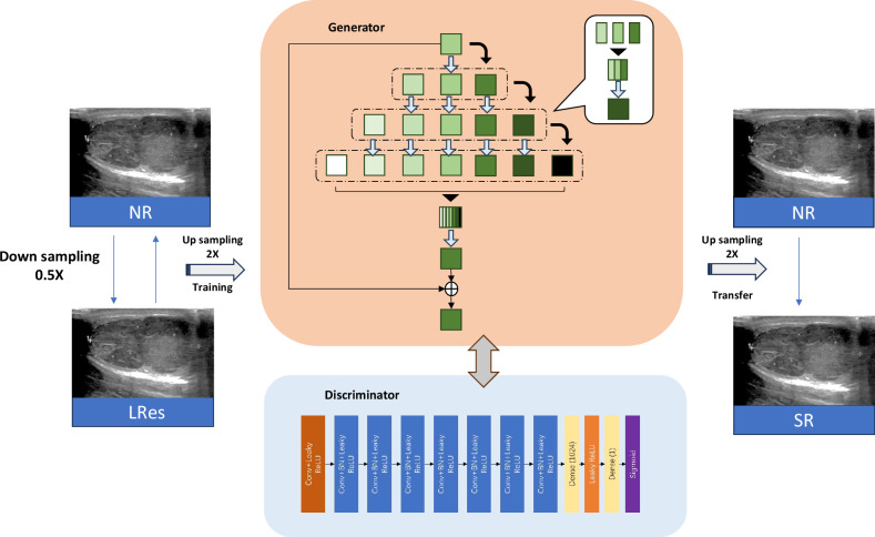
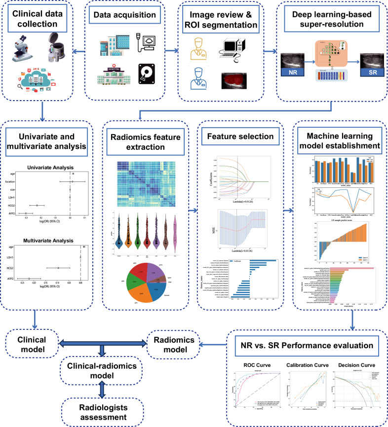
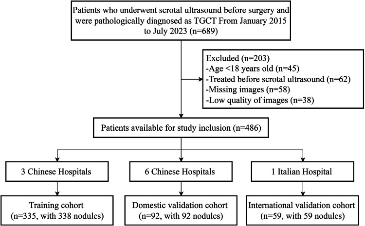
Materials and methods: This international multicenter retrospective study recruited patients with confirmed TGCT between 2015 and 2023. A pre-trained SR reconstruction algorithm was applied to enhance native resolution (NR) images. NR and SR radiomics models were constructed, and the superior model was then integrated with clinical features to construct clinical-radiomics models. Diagnostic performance was evaluated by ROC analysis (AUC) and compared with radiologists' assessments using the DeLong test.
Results: A total of 486 male patients were enrolled for training (n = 338), domestic (n = 92), and international (n = 59) validation sets. The SR radiomics model achieved AUCs of 0.90, 0.82, and 0.91, respectively, in the training, domestic, and international validation sets, significantly surpassing the NR model (p < 0.001, p = 0.031, and p = 0.001, respectively). The clinical-radiomics model exhibited a significantly higher across both domestic and international validation sets compared to the SR radiomics model alone (0.95 vs 0.82, p = 0.004; 0.97 vs 0.91, p = 0.031). Moreover, the clinical-radiomics model surpassed the performance of experienced radiologists in both domestic (AUC, 0.95 vs 0.85, p = 0.012) and international (AUC, 0.97 vs 0.77, p < 0.001) validation cohorts.
Conclusions: The SR-based clinical-radiomics model can effectively differentiate between SE and n-SE.
Critical relevance statement: This international multicenter study demonstrated that a radiomics model of deep learning-based SR reconstructed US images enabled effective differentiation between SE and n-SE.
Key points: Clinical parameters and radiologists' assessments exhibit limited diagnostic accuracy for SE/n-SE differentiation in TGCT. Based on scrotal US images of TGCT, the SR radiomics models performed better than the NR radiomics models. The SR-based clinical-radiomics model outperforms both the radiomics model and radiologists' assessment, enabling accurate, non-invasive preoperative differentiation between SE and n-SE.
期刊介绍:
Insights into Imaging (I³) is a peer-reviewed open access journal published under the brand SpringerOpen. All content published in the journal is freely available online to anyone, anywhere!
I³ continuously updates scientific knowledge and progress in best-practice standards in radiology through the publication of original articles and state-of-the-art reviews and opinions, along with recommendations and statements from the leading radiological societies in Europe.
Founded by the European Society of Radiology (ESR), I³ creates a platform for educational material, guidelines and recommendations, and a forum for topics of controversy.
A balanced combination of review articles, original papers, short communications from European radiological congresses and information on society matters makes I³ an indispensable source for current information in this field.
I³ is owned by the ESR, however authors retain copyright to their article according to the Creative Commons Attribution License (see Copyright and License Agreement). All articles can be read, redistributed and reused for free, as long as the author of the original work is cited properly.
The open access fees (article-processing charges) for this journal are kindly sponsored by ESR for all Members.
The journal went open access in 2012, which means that all articles published since then are freely available online.

 求助内容:
求助内容: 应助结果提醒方式:


