A novel method to simulate radiographs of 3D printed objects
Abstract
Background
3D printing has a number of applications within medicine and healthcare. In applications involving radiography, the internal infill structure and external geometry of a 3D printed part can produce undesirable artifacts, limiting the full potential of 3D printing as a manufacturing technology. While the mechanical performance of a 3D printed part can be easily simulated, it is difficult to simulate the radiographic artifact produced.
Purpose
The purpose of this work was to develop a tool that allows users to simulate the radiographic artifact produced by a 3D printed object.
Methods
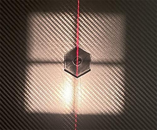
Three regular hexagons of identical geometry were sliced and 3D printed using polylactic acid (PLA) filament on a fused deposition modeling (FDM) 3D printer with varying infill patterns: rectilinear grid, cubic, and gyroid. The hexagons were then radiographed using clinical-standard scanning protocols. The captured radiographs were compared to simulated radiographs generated using the G-Code developed when the objects were sliced. The physical and simulated virtual radiographs were compared to one another, and the simulated angle of least and greatest artifact was noted.
Results
Strong visual agreement was found between the physically captured and simulated virtual radiographs. The projection angles that produced the least amount of artifact were 22.5°, 22.5°, and 12.25° for grid, cubic, and gyroid infills, respectively. The projection angles that produced the greatest amount of artifact were 0°, 45°, and 45° for grid, cubic, and gyroid infills, respectively.
Conclusions
This work provides designers of 3D printed components with a new way to evaluate a design's radiographic performance. Previously, designers would have to physically print and radiograph a part to determine the artifact produced. This work outlines the development of a tool that simulates the radiograph of a 3D printed part from multiple different projections, saving designers time to iterate to their final design.


 求助内容:
求助内容: 应助结果提醒方式:


