Predictive value of mitotic figure counts in tumor progression of non-invasive high-grade papillary urothelial carcinoma of the urinary bladder: A retrospective study from a single cancer center.
Yan Hu, Susan Karki, Weiwei Chen, Yunguang Liu, Norbert Sule, Bo Xu
{"title":"Predictive value of mitotic figure counts in tumor progression of non-invasive high-grade papillary urothelial carcinoma of the urinary bladder: A retrospective study from a single cancer center.","authors":"Yan Hu, Susan Karki, Weiwei Chen, Yunguang Liu, Norbert Sule, Bo Xu","doi":"10.14440/bladder.2024.0021","DOIUrl":null,"url":null,"abstract":"<p><strong>Background: </strong>Urothelial carcinoma (UC) is the most common type of bladder malignancy. Although the majority of UC present as non-invasive tumors, a subset of them progress into invasive cancer and cause significant morbidity and mortality.</p><p><strong>Objective: </strong>In this study, we examined the association between tumor mitotic activity associated and the progression of non-invasive high-grade papillary UC of the bladder.</p><p><strong>Methods: </strong>Forty-four cases of tumors that met the selection criteria were retrieved from the Department of Pathology archives, and, for each case, mitotic figures were counted in 10 high-power fields (HPF) by two independent pathologists. Tumor progression was defined as the invasion of the tumor into the subepithelial connective tissue (lamina propria) or beyond during follow-ups. In addition, tumors that later exhibited distant metastases were included in the tumor progression group.</p><p><strong>Results: </strong>Our study revealed that the average mitotic count per 10 HPF in the tumor progression group was significantly higher (<i>p =</i> 0.001) than in the progression-free group. Furthermore, tumors with more than three mitotic counts per HPF in initial biopsies posed a high risk of tumor progression within the 19.5 ± 6.1 months of follow-ups.</p><p><strong>Conclusion: </strong>The findings of our study provided valuable information for further stratification of risk factors among patients with non-invasive high-grade papillary UC of the bladder. Patients with high mitotic figure count in their initial biopsies should be monitored closely or treated earlier to prevent their tumors from progressing into invasive carcinoma.</p>","PeriodicalId":72421,"journal":{"name":"Bladder (San Francisco, Calif.)","volume":"12 1","pages":"e21200026"},"PeriodicalIF":0.0000,"publicationDate":"2025-01-27","publicationTypes":"Journal Article","fieldsOfStudy":null,"isOpenAccess":false,"openAccessPdf":"https://www.ncbi.nlm.nih.gov/pmc/articles/PMC12308121/pdf/","citationCount":"0","resultStr":null,"platform":"Semanticscholar","paperid":null,"PeriodicalName":"Bladder (San Francisco, Calif.)","FirstCategoryId":"1085","ListUrlMain":"https://doi.org/10.14440/bladder.2024.0021","RegionNum":0,"RegionCategory":null,"ArticlePicture":[],"TitleCN":null,"AbstractTextCN":null,"PMCID":null,"EPubDate":"2025/1/1 0:00:00","PubModel":"eCollection","JCR":"","JCRName":"","Score":null,"Total":0}
引用次数: 0
Abstract
Background: Urothelial carcinoma (UC) is the most common type of bladder malignancy. Although the majority of UC present as non-invasive tumors, a subset of them progress into invasive cancer and cause significant morbidity and mortality.
Objective: In this study, we examined the association between tumor mitotic activity associated and the progression of non-invasive high-grade papillary UC of the bladder.
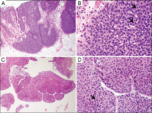
Methods: Forty-four cases of tumors that met the selection criteria were retrieved from the Department of Pathology archives, and, for each case, mitotic figures were counted in 10 high-power fields (HPF) by two independent pathologists. Tumor progression was defined as the invasion of the tumor into the subepithelial connective tissue (lamina propria) or beyond during follow-ups. In addition, tumors that later exhibited distant metastases were included in the tumor progression group.
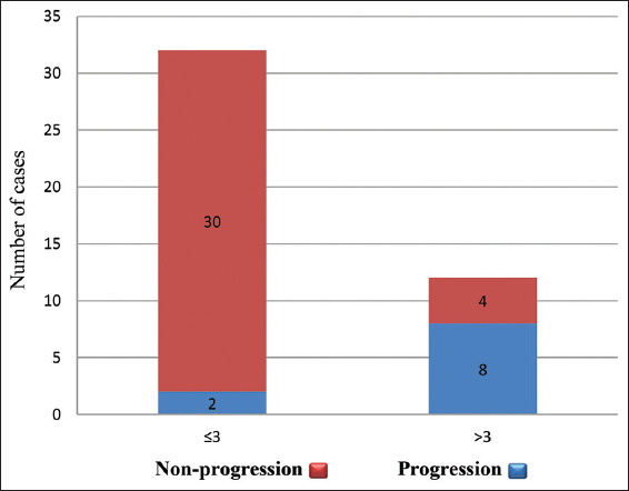
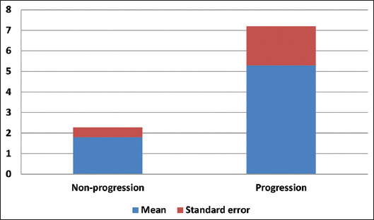
Results: Our study revealed that the average mitotic count per 10 HPF in the tumor progression group was significantly higher (p = 0.001) than in the progression-free group. Furthermore, tumors with more than three mitotic counts per HPF in initial biopsies posed a high risk of tumor progression within the 19.5 ± 6.1 months of follow-ups.
Conclusion: The findings of our study provided valuable information for further stratification of risk factors among patients with non-invasive high-grade papillary UC of the bladder. Patients with high mitotic figure count in their initial biopsies should be monitored closely or treated earlier to prevent their tumors from progressing into invasive carcinoma.

 求助内容:
求助内容: 应助结果提醒方式:


