Quantifying the Trajectory of Percutaneous Endoscopic Lumbar Discectomy in 3D Lumbar Models Based on Automated MR Image Segmentation-A Cross-Sectional Study.
Zhihai Su, Yunfei Wang, Chengjie Huang, Qingqing He, Junjie Lu, Zheng Liu, Yiou Zhang, Qiaochu Zhao, YuChen Zhang, Jianan Cai, Shumao Pang, Zhen Yuan, Ziyang Chen, Tao Chen, Hai Lu
{"title":"Quantifying the Trajectory of Percutaneous Endoscopic Lumbar Discectomy in 3D Lumbar Models Based on Automated MR Image Segmentation-A Cross-Sectional Study.","authors":"Zhihai Su, Yunfei Wang, Chengjie Huang, Qingqing He, Junjie Lu, Zheng Liu, Yiou Zhang, Qiaochu Zhao, YuChen Zhang, Jianan Cai, Shumao Pang, Zhen Yuan, Ziyang Chen, Tao Chen, Hai Lu","doi":"10.1111/os.70112","DOIUrl":null,"url":null,"abstract":"<p><strong>Objective: </strong>Creating a 3D lumbar model and planning a personalized puncture trajectory has an advantage in establishing the working channel for percutaneous endoscopic lumbar discectomy (PELD). However, existing 3D lumbar models, which seldom include lumbar nerves and dural sac reconstructions, primarily depend on CT images for preoperative trajectory planning. Therefore, our study aims to further investigate the relationship between different virtual working channels and the 3D lumbar model, which includes automated MR image segmentation of lumbar bone, nerves, and dural sac at the L4/L5 level.</p><p><strong>Methods: </strong>Preoperative lumbar MR images of 50 patients with L4/L5 lumbar disc herniation were collected from a teaching hospital between March 2020 and July 2020. Automated MR image segmentation was initially used to create a 3D model of the lumbar spine, including the L4 vertebrae, L5 vertebrae, intervertebral disc, L4 nerves, dural sac, and skin. Thirty were then randomly chosen from the segmentation results to clarify the relationship between various virtual working channels and the lumbar 3D model. A bivariate Spearman's rank correlation analysis was used in this study.</p><p><strong>Results: </strong>Preoperative MR images of 50 patients (34 males, mean age 45.6 ± 6 years) were used to train and validate the automated segmentation model, which had mean Dice scores of 0.906, 0.891, 0.896, 0.695, 0.892, and 0.892 for the L4 vertebrae, L5 vertebrae, intervertebral disc, L4 nerves, dural sac, and skin, respectively. With an increase in the coronal plane angle (CPA), there was a reduction in the intersection volume involving the L4 nerves and atypical structures. Conversely, the intersection volume encompassing the dural sac, L4 inferior articular process, and L5 superior articular process increased; the total intersection volume showed a fluctuating pattern: it initially decreased, followed by an increase, and then decreased once more. As the cross-section angle (CSA) increased, there was a rise in the intersection volume of both the L4 nerves and the dural sac; the intersection volume involving the L4 inferior articular process grew while that of the L5 superior articular process diminished; the overall intersection volume and the intersection volume of atypical structures initially decreased, followed by an increase.</p><p><strong>Conclusion: </strong>In terms of regularity, the optimal angles for L4/L5 PELD are a CSA of 15° and a CPA of 15°-20°, minimizing harm to the vertebral bones, facet joint, spinal nerves, and dural sac. Additionally, our 3D preoperative planning method could enhance puncture trajectories for individual patients, potentially advancing surgical navigation, robots, and artificial intelligence in PELD procedures.</p>","PeriodicalId":19566,"journal":{"name":"Orthopaedic Surgery","volume":" ","pages":"2689-2698"},"PeriodicalIF":2.1000,"publicationDate":"2025-09-01","publicationTypes":"Journal Article","fieldsOfStudy":null,"isOpenAccess":false,"openAccessPdf":"https://www.ncbi.nlm.nih.gov/pmc/articles/PMC12404874/pdf/","citationCount":"0","resultStr":null,"platform":"Semanticscholar","paperid":null,"PeriodicalName":"Orthopaedic Surgery","FirstCategoryId":"3","ListUrlMain":"https://doi.org/10.1111/os.70112","RegionNum":2,"RegionCategory":"医学","ArticlePicture":[],"TitleCN":null,"AbstractTextCN":null,"PMCID":null,"EPubDate":"2025/7/31 0:00:00","PubModel":"Epub","JCR":"Q2","JCRName":"ORTHOPEDICS","Score":null,"Total":0}
引用次数: 0
Abstract
Objective: Creating a 3D lumbar model and planning a personalized puncture trajectory has an advantage in establishing the working channel for percutaneous endoscopic lumbar discectomy (PELD). However, existing 3D lumbar models, which seldom include lumbar nerves and dural sac reconstructions, primarily depend on CT images for preoperative trajectory planning. Therefore, our study aims to further investigate the relationship between different virtual working channels and the 3D lumbar model, which includes automated MR image segmentation of lumbar bone, nerves, and dural sac at the L4/L5 level.
Methods: Preoperative lumbar MR images of 50 patients with L4/L5 lumbar disc herniation were collected from a teaching hospital between March 2020 and July 2020. Automated MR image segmentation was initially used to create a 3D model of the lumbar spine, including the L4 vertebrae, L5 vertebrae, intervertebral disc, L4 nerves, dural sac, and skin. Thirty were then randomly chosen from the segmentation results to clarify the relationship between various virtual working channels and the lumbar 3D model. A bivariate Spearman's rank correlation analysis was used in this study.
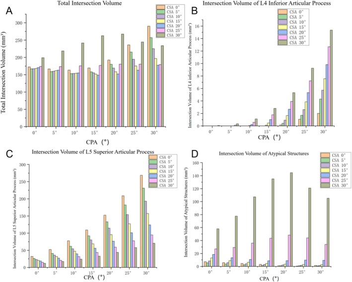
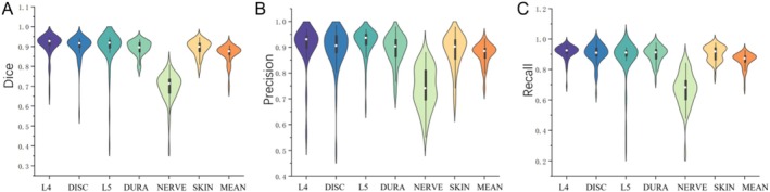
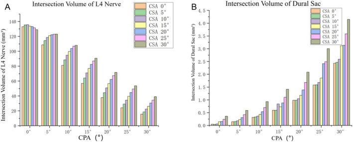
Results: Preoperative MR images of 50 patients (34 males, mean age 45.6 ± 6 years) were used to train and validate the automated segmentation model, which had mean Dice scores of 0.906, 0.891, 0.896, 0.695, 0.892, and 0.892 for the L4 vertebrae, L5 vertebrae, intervertebral disc, L4 nerves, dural sac, and skin, respectively. With an increase in the coronal plane angle (CPA), there was a reduction in the intersection volume involving the L4 nerves and atypical structures. Conversely, the intersection volume encompassing the dural sac, L4 inferior articular process, and L5 superior articular process increased; the total intersection volume showed a fluctuating pattern: it initially decreased, followed by an increase, and then decreased once more. As the cross-section angle (CSA) increased, there was a rise in the intersection volume of both the L4 nerves and the dural sac; the intersection volume involving the L4 inferior articular process grew while that of the L5 superior articular process diminished; the overall intersection volume and the intersection volume of atypical structures initially decreased, followed by an increase.
Conclusion: In terms of regularity, the optimal angles for L4/L5 PELD are a CSA of 15° and a CPA of 15°-20°, minimizing harm to the vertebral bones, facet joint, spinal nerves, and dural sac. Additionally, our 3D preoperative planning method could enhance puncture trajectories for individual patients, potentially advancing surgical navigation, robots, and artificial intelligence in PELD procedures.
期刊介绍:
Orthopaedic Surgery (OS) is the official journal of the Chinese Orthopaedic Association, focusing on all aspects of orthopaedic technique and surgery.
The journal publishes peer-reviewed articles in the following categories: Original Articles, Clinical Articles, Review Articles, Guidelines, Editorials, Commentaries, Surgical Techniques, Case Reports and Meeting Reports.

 求助内容:
求助内容: 应助结果提醒方式:


