{"title":"Establishment of Subtrochanteric Fracture in Pre-Clinical Animal Model.","authors":"Poornima Palanisamy, Simon Kwoon-Ho Chow, Shuai Li, Michelle Meng-Chen Li, Wing-Hoi Cheung, Ling Qin, Yong-Ping Zheng","doi":"10.1111/os.70137","DOIUrl":null,"url":null,"abstract":"<p><strong>Objective: </strong>About 7%-34% of the femur fracture contributes to subtrochanteric fracture, and only very little research is available about these fractures when compared to common hip fractures. Hence, the aim of this study was to develop a clinically relevant and reproducible open fracture model in rabbits at the subtrochanteric region to understand the fracture healing mechanism at this site and to explore treatment effects of biophysical intervention in future studies.</p><p><strong>Methods: </strong>An open osteotomy was created in 32 adult New Zealand white rabbits at the subtrochanteric region, followed by customized titanium internal fixations. The internal fixator consists of a 3D printed titanium compression plate with cortical screws for locking. The fracture healing was monitored for 6 weeks, and the corresponding radiography, MicroCT, and histomorphometry analysis were performed at regular intervals.</p><p><strong>Results: </strong>Four rabbits were excluded due to complications (4/32), including bone dislocation one week post-surgery (3/32). Fracture healing progression was observed in radiographic images. MicroCT analysis showed increased callus volume after 42 days. Histomorphometry revealed remodeled bone area with a higher number of osteocyte cells.</p><p><strong>Conclusion: </strong>The rabbit fracture model of an open femoral osteotomy at the subtrochanteric region has been successfully established, with the facilitation of an internal fixator consisting of a 3D printed titanium compression plate with cortical screws for locking. Applications of this model are being investigated, including different biophysical stimulation methods for accelerating fracture healing.</p>","PeriodicalId":19566,"journal":{"name":"Orthopaedic Surgery","volume":" ","pages":"2726-2734"},"PeriodicalIF":2.1000,"publicationDate":"2025-09-01","publicationTypes":"Journal Article","fieldsOfStudy":null,"isOpenAccess":false,"openAccessPdf":"https://www.ncbi.nlm.nih.gov/pmc/articles/PMC12404857/pdf/","citationCount":"0","resultStr":null,"platform":"Semanticscholar","paperid":null,"PeriodicalName":"Orthopaedic Surgery","FirstCategoryId":"3","ListUrlMain":"https://doi.org/10.1111/os.70137","RegionNum":2,"RegionCategory":"医学","ArticlePicture":[],"TitleCN":null,"AbstractTextCN":null,"PMCID":null,"EPubDate":"2025/7/31 0:00:00","PubModel":"Epub","JCR":"Q2","JCRName":"ORTHOPEDICS","Score":null,"Total":0}
引用次数: 0
Abstract
Objective: About 7%-34% of the femur fracture contributes to subtrochanteric fracture, and only very little research is available about these fractures when compared to common hip fractures. Hence, the aim of this study was to develop a clinically relevant and reproducible open fracture model in rabbits at the subtrochanteric region to understand the fracture healing mechanism at this site and to explore treatment effects of biophysical intervention in future studies.
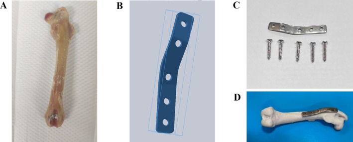
Methods: An open osteotomy was created in 32 adult New Zealand white rabbits at the subtrochanteric region, followed by customized titanium internal fixations. The internal fixator consists of a 3D printed titanium compression plate with cortical screws for locking. The fracture healing was monitored for 6 weeks, and the corresponding radiography, MicroCT, and histomorphometry analysis were performed at regular intervals.
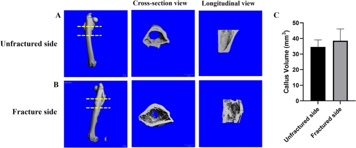
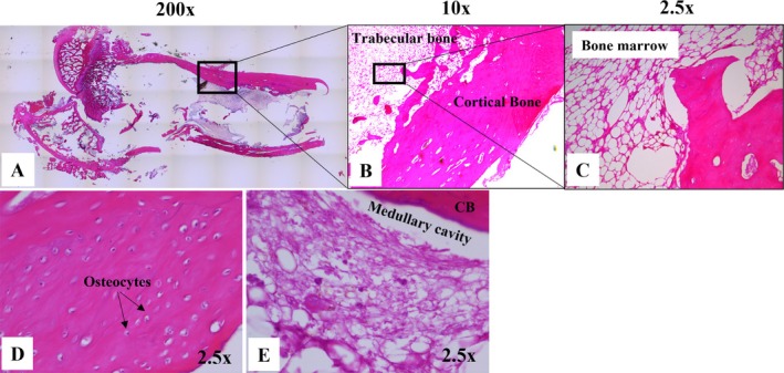
Results: Four rabbits were excluded due to complications (4/32), including bone dislocation one week post-surgery (3/32). Fracture healing progression was observed in radiographic images. MicroCT analysis showed increased callus volume after 42 days. Histomorphometry revealed remodeled bone area with a higher number of osteocyte cells.
Conclusion: The rabbit fracture model of an open femoral osteotomy at the subtrochanteric region has been successfully established, with the facilitation of an internal fixator consisting of a 3D printed titanium compression plate with cortical screws for locking. Applications of this model are being investigated, including different biophysical stimulation methods for accelerating fracture healing.
期刊介绍:
Orthopaedic Surgery (OS) is the official journal of the Chinese Orthopaedic Association, focusing on all aspects of orthopaedic technique and surgery.
The journal publishes peer-reviewed articles in the following categories: Original Articles, Clinical Articles, Review Articles, Guidelines, Editorials, Commentaries, Surgical Techniques, Case Reports and Meeting Reports.
 求助内容:
求助内容: 应助结果提醒方式:


