{"title":"The value of a radiomics model in predicting ovarian malignancy: a retrospective multi-center comparison with O-RADS and radiologists.","authors":"Junjie Jin, Xijia Deng, Ling Long, Meiling Liu, Meimei Cao, Hao Gong, Huan Liu, Xiaosong Lan, Lili Liu, Jiuquan Zhang","doi":"10.1186/s13244-025-02047-w","DOIUrl":null,"url":null,"abstract":"<p><strong>Objectives: </strong>To develop an MRI-based radiomics model for ovarian masses categorization and to compare the model performance to Ovarian-Adnexal Reporting and Data System (O-RADS) and radiologists' assessments.</p><p><strong>Materials and methods: </strong>This retrospective multicenter study included 497 patients (249 benign, 248 malignant) allocated to training, internal, and external validation sets (293/124/80 masses, respectively). Radiomics features were extracted from preoperative MRI. Features were selected using minimum redundancy, maximum relevance, and the least absolute shrinkage and selection operator algorithm. Diagnostic performance of the radiomics model, O-RADS, and independent assessments by junior and senior radiologists was evaluated via the area under the receiver operating characteristic curve (AUC) and compared using DeLong's test.</p><p><strong>Results: </strong>In external validation, the radiomics model (AUC = 0.939) outperformed O-RADS (AUC = 0.862; p = 0.047) and the junior radiologist (AUC = 0.802; p = 0.003) and was similar to the senior radiologist (AUC = 0.886; p = 0.231). Subgroup analysis of O-RADS score 4 showed the model (AUC = 0.879) outperformed both radiologists (junior: p = 0.001; senior: p = 0.005). For solid, cystic-solids, and cystic masses, the AUCs of the model were 0.921, 0.975, and 0.848, respectively.</p><p><strong>Conclusions: </strong>The performance of the radiomics model to categorize ovarian masses was superior to O-RADS and junior radiologists and similar to senior radiologists. As a complementary tool to O-RADS, it allows for refined risk stratification for ovarian masses with an O-RADS score of 4 and different morphological characteristics, providing clinicians with quantitative decision support to improve preoperative diagnosis and guide treatment planning.</p><p><strong>Critical relevance statement: </strong>Radiomics model provides improved risk stratification and supports precise clinical decision-making for ovarian masses with an O-RADS score of 4 and solid, cystic-solid ovarian masses, thereby improving the management of patients with ovarian masses.</p><p><strong>Key points: </strong>MRI-based radiomics allows for the characterization of ovarian masses with high accuracy. Radiomics helps differentiate between benign and malignant ovarian masses with an O-RADS score of 4. For solid, cystic-solid, and cystic masses, the radiomics model exhibited higher or similar performance to that of the O-RADS and radiologists.</p>","PeriodicalId":13639,"journal":{"name":"Insights into Imaging","volume":"16 1","pages":"163"},"PeriodicalIF":4.5000,"publicationDate":"2025-07-31","publicationTypes":"Journal Article","fieldsOfStudy":null,"isOpenAccess":false,"openAccessPdf":"https://www.ncbi.nlm.nih.gov/pmc/articles/PMC12314133/pdf/","citationCount":"0","resultStr":null,"platform":"Semanticscholar","paperid":null,"PeriodicalName":"Insights into Imaging","FirstCategoryId":"3","ListUrlMain":"https://doi.org/10.1186/s13244-025-02047-w","RegionNum":2,"RegionCategory":"医学","ArticlePicture":[],"TitleCN":null,"AbstractTextCN":null,"PMCID":null,"EPubDate":"","PubModel":"","JCR":"Q1","JCRName":"RADIOLOGY, NUCLEAR MEDICINE & MEDICAL IMAGING","Score":null,"Total":0}
引用次数: 0
Abstract
Objectives: To develop an MRI-based radiomics model for ovarian masses categorization and to compare the model performance to Ovarian-Adnexal Reporting and Data System (O-RADS) and radiologists' assessments.
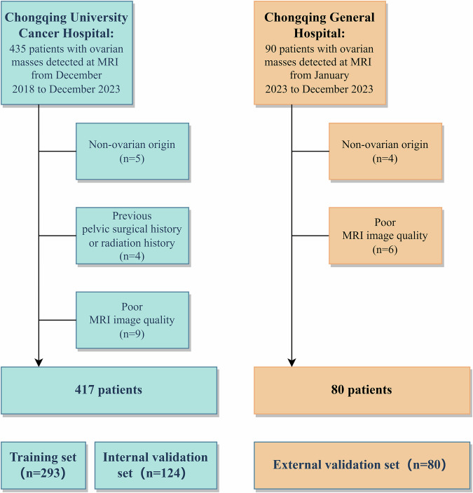
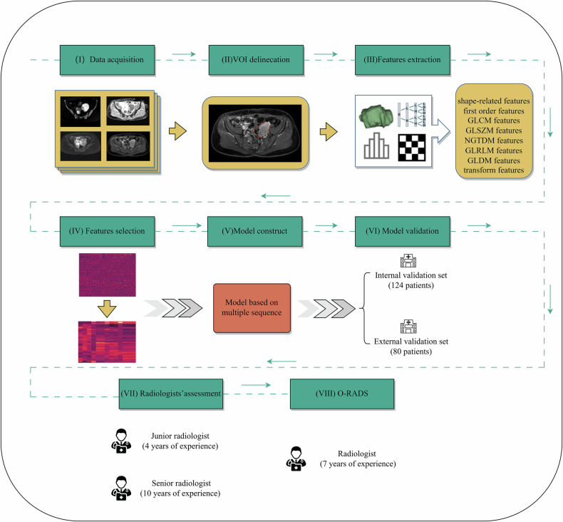
Materials and methods: This retrospective multicenter study included 497 patients (249 benign, 248 malignant) allocated to training, internal, and external validation sets (293/124/80 masses, respectively). Radiomics features were extracted from preoperative MRI. Features were selected using minimum redundancy, maximum relevance, and the least absolute shrinkage and selection operator algorithm. Diagnostic performance of the radiomics model, O-RADS, and independent assessments by junior and senior radiologists was evaluated via the area under the receiver operating characteristic curve (AUC) and compared using DeLong's test.
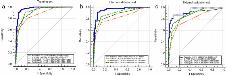
Results: In external validation, the radiomics model (AUC = 0.939) outperformed O-RADS (AUC = 0.862; p = 0.047) and the junior radiologist (AUC = 0.802; p = 0.003) and was similar to the senior radiologist (AUC = 0.886; p = 0.231). Subgroup analysis of O-RADS score 4 showed the model (AUC = 0.879) outperformed both radiologists (junior: p = 0.001; senior: p = 0.005). For solid, cystic-solids, and cystic masses, the AUCs of the model were 0.921, 0.975, and 0.848, respectively.
Conclusions: The performance of the radiomics model to categorize ovarian masses was superior to O-RADS and junior radiologists and similar to senior radiologists. As a complementary tool to O-RADS, it allows for refined risk stratification for ovarian masses with an O-RADS score of 4 and different morphological characteristics, providing clinicians with quantitative decision support to improve preoperative diagnosis and guide treatment planning.
Critical relevance statement: Radiomics model provides improved risk stratification and supports precise clinical decision-making for ovarian masses with an O-RADS score of 4 and solid, cystic-solid ovarian masses, thereby improving the management of patients with ovarian masses.
Key points: MRI-based radiomics allows for the characterization of ovarian masses with high accuracy. Radiomics helps differentiate between benign and malignant ovarian masses with an O-RADS score of 4. For solid, cystic-solid, and cystic masses, the radiomics model exhibited higher or similar performance to that of the O-RADS and radiologists.
期刊介绍:
Insights into Imaging (I³) is a peer-reviewed open access journal published under the brand SpringerOpen. All content published in the journal is freely available online to anyone, anywhere!
I³ continuously updates scientific knowledge and progress in best-practice standards in radiology through the publication of original articles and state-of-the-art reviews and opinions, along with recommendations and statements from the leading radiological societies in Europe.
Founded by the European Society of Radiology (ESR), I³ creates a platform for educational material, guidelines and recommendations, and a forum for topics of controversy.
A balanced combination of review articles, original papers, short communications from European radiological congresses and information on society matters makes I³ an indispensable source for current information in this field.
I³ is owned by the ESR, however authors retain copyright to their article according to the Creative Commons Attribution License (see Copyright and License Agreement). All articles can be read, redistributed and reused for free, as long as the author of the original work is cited properly.
The open access fees (article-processing charges) for this journal are kindly sponsored by ESR for all Members.
The journal went open access in 2012, which means that all articles published since then are freely available online.

 求助内容:
求助内容: 应助结果提醒方式:


