Ziqi Wu, Liya Gong, Jingwen Luo, Xiaobo Chen, Fan Yang, Junyan Wen, Yanyu Hao, Zhishan Wang, Ruozhen Gu, Yuqin Zhang, Hai Liao, Ge Wen
{"title":"An interpretable CT-based machine learning model for predicting recurrence risk in stage II colorectal cancer.","authors":"Ziqi Wu, Liya Gong, Jingwen Luo, Xiaobo Chen, Fan Yang, Junyan Wen, Yanyu Hao, Zhishan Wang, Ruozhen Gu, Yuqin Zhang, Hai Liao, Ge Wen","doi":"10.1186/s13244-025-02009-2","DOIUrl":null,"url":null,"abstract":"<p><strong>Objectives: </strong>This study aimed to develop an interpretable 3-year disease-free survival risk prediction tool to stratify patients with stage II colorectal cancer (CRC) by integrating CT images and clinicopathological factors.</p><p><strong>Methods: </strong>A total of 769 patients with pathologically confirmed stage II CRC and disease-free survival (DFS) follow-up information were recruited from three medical centers and divided into training (n = 442), test (n = 190), and validation cohorts (n = 137). CT-based tumor radiomics features were extracted, selected, and used to calculate a Radscore. A combined model was developed using artificial neural network (ANN) algorithm, by integrating the Radscore with significant clinicoradiological factors to classify patients into high- and low-risk groups. Model performance was assessed using the area under the curve (AUC), and feature contributions were qualified using the Shapley additive explanation (SHAP) algorithm. Kaplan-Meier survival analysis revealed the prognostic stratification value of the risk groups.</p><p><strong>Results: </strong>Fourteen radiomics features and five clinicoradiological factors were selected to construct the radiomics and clinicoradiological models, respectively. The combined model demonstrated optimal performance, with AUCs of 0.811 and 0.846 in the test and validation cohorts, respectively. Kaplan-Meier curves confirmed effective patient stratification (p < 0.001) in both test and validation cohorts. A high Radscore, rough intestinal outer edge, and advanced age were identified as key prognostic risk factors using the SHAP.</p><p><strong>Conclusion: </strong>The combined model effectively stratified patients with stage II CRC into different prognostic risk groups, aiding clinical decision-making.</p><p><strong>Critical relevance statement: </strong>Integrating CT images with clinicopathological information can facilitate the identification of patients with stage II CRC who are most likely to benefit from adjuvant chemotherapy.</p><p><strong>Key points: </strong>The effectiveness of adjuvant chemotherapy for stage II colorectal cancer remains debated. A combined model successfully identified high-risk stage II colorectal cancer patients. Shapley additive explanations enhance the interpretability of the model's predictions.</p>","PeriodicalId":13639,"journal":{"name":"Insights into Imaging","volume":"16 1","pages":"162"},"PeriodicalIF":4.5000,"publicationDate":"2025-07-31","publicationTypes":"Journal Article","fieldsOfStudy":null,"isOpenAccess":false,"openAccessPdf":"https://www.ncbi.nlm.nih.gov/pmc/articles/PMC12314294/pdf/","citationCount":"0","resultStr":null,"platform":"Semanticscholar","paperid":null,"PeriodicalName":"Insights into Imaging","FirstCategoryId":"3","ListUrlMain":"https://doi.org/10.1186/s13244-025-02009-2","RegionNum":2,"RegionCategory":"医学","ArticlePicture":[],"TitleCN":null,"AbstractTextCN":null,"PMCID":null,"EPubDate":"","PubModel":"","JCR":"Q1","JCRName":"RADIOLOGY, NUCLEAR MEDICINE & MEDICAL IMAGING","Score":null,"Total":0}
引用次数: 0
Abstract
Objectives: This study aimed to develop an interpretable 3-year disease-free survival risk prediction tool to stratify patients with stage II colorectal cancer (CRC) by integrating CT images and clinicopathological factors.
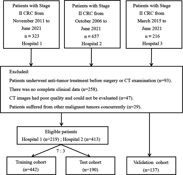
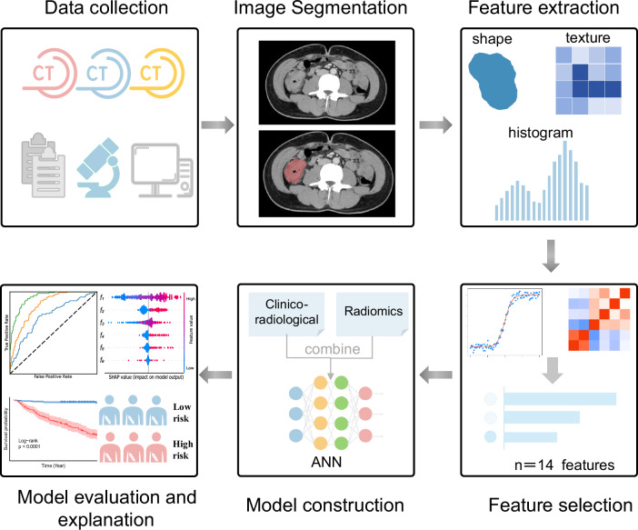
Methods: A total of 769 patients with pathologically confirmed stage II CRC and disease-free survival (DFS) follow-up information were recruited from three medical centers and divided into training (n = 442), test (n = 190), and validation cohorts (n = 137). CT-based tumor radiomics features were extracted, selected, and used to calculate a Radscore. A combined model was developed using artificial neural network (ANN) algorithm, by integrating the Radscore with significant clinicoradiological factors to classify patients into high- and low-risk groups. Model performance was assessed using the area under the curve (AUC), and feature contributions were qualified using the Shapley additive explanation (SHAP) algorithm. Kaplan-Meier survival analysis revealed the prognostic stratification value of the risk groups.
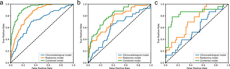
Results: Fourteen radiomics features and five clinicoradiological factors were selected to construct the radiomics and clinicoradiological models, respectively. The combined model demonstrated optimal performance, with AUCs of 0.811 and 0.846 in the test and validation cohorts, respectively. Kaplan-Meier curves confirmed effective patient stratification (p < 0.001) in both test and validation cohorts. A high Radscore, rough intestinal outer edge, and advanced age were identified as key prognostic risk factors using the SHAP.
Conclusion: The combined model effectively stratified patients with stage II CRC into different prognostic risk groups, aiding clinical decision-making.
Critical relevance statement: Integrating CT images with clinicopathological information can facilitate the identification of patients with stage II CRC who are most likely to benefit from adjuvant chemotherapy.
Key points: The effectiveness of adjuvant chemotherapy for stage II colorectal cancer remains debated. A combined model successfully identified high-risk stage II colorectal cancer patients. Shapley additive explanations enhance the interpretability of the model's predictions.
期刊介绍:
Insights into Imaging (I³) is a peer-reviewed open access journal published under the brand SpringerOpen. All content published in the journal is freely available online to anyone, anywhere!
I³ continuously updates scientific knowledge and progress in best-practice standards in radiology through the publication of original articles and state-of-the-art reviews and opinions, along with recommendations and statements from the leading radiological societies in Europe.
Founded by the European Society of Radiology (ESR), I³ creates a platform for educational material, guidelines and recommendations, and a forum for topics of controversy.
A balanced combination of review articles, original papers, short communications from European radiological congresses and information on society matters makes I³ an indispensable source for current information in this field.
I³ is owned by the ESR, however authors retain copyright to their article according to the Creative Commons Attribution License (see Copyright and License Agreement). All articles can be read, redistributed and reused for free, as long as the author of the original work is cited properly.
The open access fees (article-processing charges) for this journal are kindly sponsored by ESR for all Members.
The journal went open access in 2012, which means that all articles published since then are freely available online.

 求助内容:
求助内容: 应助结果提醒方式:


