Shirley Man, Teunis B H Geijtenbeek, Neeltje A Kootstra
{"title":"Flow Cytometric Quantification of HIV-1-Infected Cells Expressing Either Abortive or Elongated HIV-1 Transcripts Using Flow-FISH.","authors":"Shirley Man, Teunis B H Geijtenbeek, Neeltje A Kootstra","doi":"10.21769/BioProtoc.5392","DOIUrl":null,"url":null,"abstract":"<p><p>The persistence of the HIV-1 reservoir remains the ultimate obstacle in achieving a cure. Cure strategies targeting the HIV-1 reservoir are under development, and therefore, finding ways to improve the detection of the reservoir is crucial. Several reservoir detection techniques exist to assess different markers of the HIV-1 reservoir, such as PCR-based assays and protein-based flow cytometric methods. We developed a flow cytometry-fluorescent in situ hybridization (flow-FISH) approach that assesses HIV-1 at the transcriptional level. Using a combination of probes that target either the HIV-1 trans-activation response (TAR) region and 5' long terminal repeat (LTR) or the Gag sequence, our assay distinguishes between infected cells expressing abortive or elongated HIV-1 RNAs. This assay utilizes the branched-DNA method to amplify the fluorescent signal of the hybridized RNA probes and can be used directly for thawed or cultured cells, with the option to include surface antibody staining. Cellular expression of abortive and/or Gag HIV-1 RNAs is measured by flow cytometry. Our flow-FISH approach gives insight into the transcriptional dynamics of the HIV-1 reservoir and allows for the characterization of latently infected cells. Key features • Detection of latently HIV-1-infected cells identified by the expression of abortive HIV-1 TAR transcripts. • Cell activation is not required for HIV-1 detection; therefore, the cellular phenotypic landscape remains preserved. • Can be used for direct ex vivo measurements in isolated cells, such as PBMCs, from untreated and antiretroviral therapy (ART)-treated people with HIV-1 (PWH).</p>","PeriodicalId":93907,"journal":{"name":"Bio-protocol","volume":"15 14","pages":"e5392"},"PeriodicalIF":1.1000,"publicationDate":"2025-07-20","publicationTypes":"Journal Article","fieldsOfStudy":null,"isOpenAccess":false,"openAccessPdf":"https://www.ncbi.nlm.nih.gov/pmc/articles/PMC12304468/pdf/","citationCount":"0","resultStr":null,"platform":"Semanticscholar","paperid":null,"PeriodicalName":"Bio-protocol","FirstCategoryId":"1085","ListUrlMain":"https://doi.org/10.21769/BioProtoc.5392","RegionNum":0,"RegionCategory":null,"ArticlePicture":[],"TitleCN":null,"AbstractTextCN":null,"PMCID":null,"EPubDate":"","PubModel":"","JCR":"Q3","JCRName":"BIOLOGY","Score":null,"Total":0}
引用次数: 0
Abstract
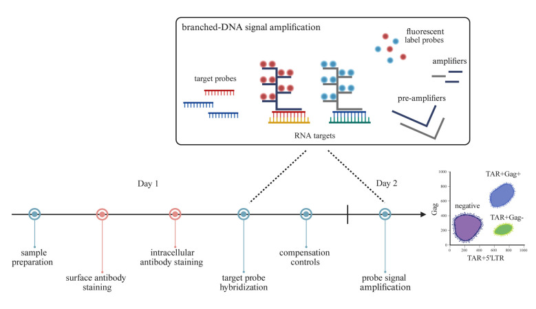
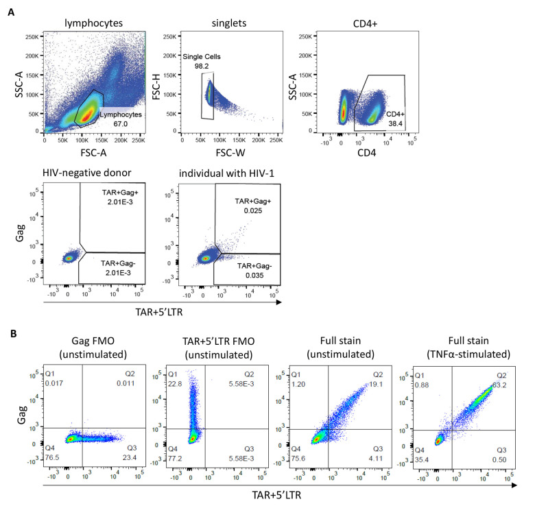
The persistence of the HIV-1 reservoir remains the ultimate obstacle in achieving a cure. Cure strategies targeting the HIV-1 reservoir are under development, and therefore, finding ways to improve the detection of the reservoir is crucial. Several reservoir detection techniques exist to assess different markers of the HIV-1 reservoir, such as PCR-based assays and protein-based flow cytometric methods. We developed a flow cytometry-fluorescent in situ hybridization (flow-FISH) approach that assesses HIV-1 at the transcriptional level. Using a combination of probes that target either the HIV-1 trans-activation response (TAR) region and 5' long terminal repeat (LTR) or the Gag sequence, our assay distinguishes between infected cells expressing abortive or elongated HIV-1 RNAs. This assay utilizes the branched-DNA method to amplify the fluorescent signal of the hybridized RNA probes and can be used directly for thawed or cultured cells, with the option to include surface antibody staining. Cellular expression of abortive and/or Gag HIV-1 RNAs is measured by flow cytometry. Our flow-FISH approach gives insight into the transcriptional dynamics of the HIV-1 reservoir and allows for the characterization of latently infected cells. Key features • Detection of latently HIV-1-infected cells identified by the expression of abortive HIV-1 TAR transcripts. • Cell activation is not required for HIV-1 detection; therefore, the cellular phenotypic landscape remains preserved. • Can be used for direct ex vivo measurements in isolated cells, such as PBMCs, from untreated and antiretroviral therapy (ART)-treated people with HIV-1 (PWH).

 求助内容:
求助内容: 应助结果提醒方式:


