{"title":"The Analysis of Magnetic Resonance Imaging on the Intra- and Extra-Osseous Blood Supply After Scaphoid Fractures.","authors":"Wei Zheng, Ge Xiong, Zhe Guo, Wei Zhang, Zongxuan Zhao, Jiangchao Zhang","doi":"10.1111/os.70136","DOIUrl":null,"url":null,"abstract":"<p><strong>Objectives: </strong>Scaphoid fractures are the most common carpal fractures with a relatively high incidence of nonunion and avascular necrosis. Previous autopsy studies have shown that proximal ischemia after a fracture is related to the fracture site and retrograde blood supply within the scaphoid, but actual changes in blood flow after fractures have not been thoroughly studied. The purpose of this study was to analyze the influencing factors of blood supply after scaphoid fractures.</p><p><strong>Methods: </strong>This was a prospective study. Between 2022 and 2023, contrast-enhanced magnetic resonance angiography and gadolinium-enhanced magnetic resonance imaging were performed in 32 patients (28 males and 4 females) with scaphoid fractures. The average age was 35 years (ranges: 15-74 years). We observed the accumulation and filling time of contrast media, and measured the diameters of extraosseous arteries as well as the signal intensity of intraosseous perfusion. The Mann-Whitney U-test, student's t-test, and Friedman test were used, respectively.</p><p><strong>Results: </strong>31 of 32 patients showed contrast media accumulation in the wrist joints on the affected sides. The filling time of contrast media on the affected sides was 5.6 (6.05-1) s quicker than on the healthy sides. The diameters of the radial arteries on the affected side increased by 12.8% (SD, 18.4%) compared to those on the healthy side (p = 0.002). All the patients had visible scaphoid nutrient arteries originating from the radial arteries on the affected side. The number of visible arteries on the healthy side was lower. Blood supply to the scaphoid was not related to the patient's sex, injury side, or fracture site. The increase in blood supply at the proximal fragment in older patients was less than that in young individuals (p = 0.015). Blood supply to the proximal and distal fragments of the scaphoid increased after fracture (p < 0.05). Within 1 month after the fracture, the increase in blood supply at the proximal fragment was less than that at the distal fragment, and it increased significantly after 1 month (p = 0.014). However, long-term nonunion (more than 4 years) leads to a decrease in proximal blood supply.</p><p><strong>Conclusions: </strong>The unique blood supply pattern of the scaphoid and fracture sites might not be the cause of ischemia after a fracture. This could be due to prolonged nonhealing, which leads to proximal ischemia.</p>","PeriodicalId":19566,"journal":{"name":"Orthopaedic Surgery","volume":" ","pages":"2617-2628"},"PeriodicalIF":2.1000,"publicationDate":"2025-09-01","publicationTypes":"Journal Article","fieldsOfStudy":null,"isOpenAccess":false,"openAccessPdf":"https://www.ncbi.nlm.nih.gov/pmc/articles/PMC12404867/pdf/","citationCount":"0","resultStr":null,"platform":"Semanticscholar","paperid":null,"PeriodicalName":"Orthopaedic Surgery","FirstCategoryId":"3","ListUrlMain":"https://doi.org/10.1111/os.70136","RegionNum":2,"RegionCategory":"医学","ArticlePicture":[],"TitleCN":null,"AbstractTextCN":null,"PMCID":null,"EPubDate":"2025/7/30 0:00:00","PubModel":"Epub","JCR":"Q2","JCRName":"ORTHOPEDICS","Score":null,"Total":0}
引用次数: 0
Abstract
Objectives: Scaphoid fractures are the most common carpal fractures with a relatively high incidence of nonunion and avascular necrosis. Previous autopsy studies have shown that proximal ischemia after a fracture is related to the fracture site and retrograde blood supply within the scaphoid, but actual changes in blood flow after fractures have not been thoroughly studied. The purpose of this study was to analyze the influencing factors of blood supply after scaphoid fractures.
Methods: This was a prospective study. Between 2022 and 2023, contrast-enhanced magnetic resonance angiography and gadolinium-enhanced magnetic resonance imaging were performed in 32 patients (28 males and 4 females) with scaphoid fractures. The average age was 35 years (ranges: 15-74 years). We observed the accumulation and filling time of contrast media, and measured the diameters of extraosseous arteries as well as the signal intensity of intraosseous perfusion. The Mann-Whitney U-test, student's t-test, and Friedman test were used, respectively.
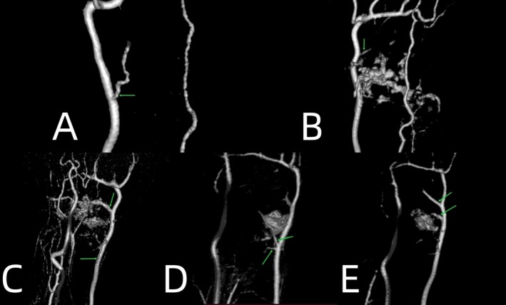
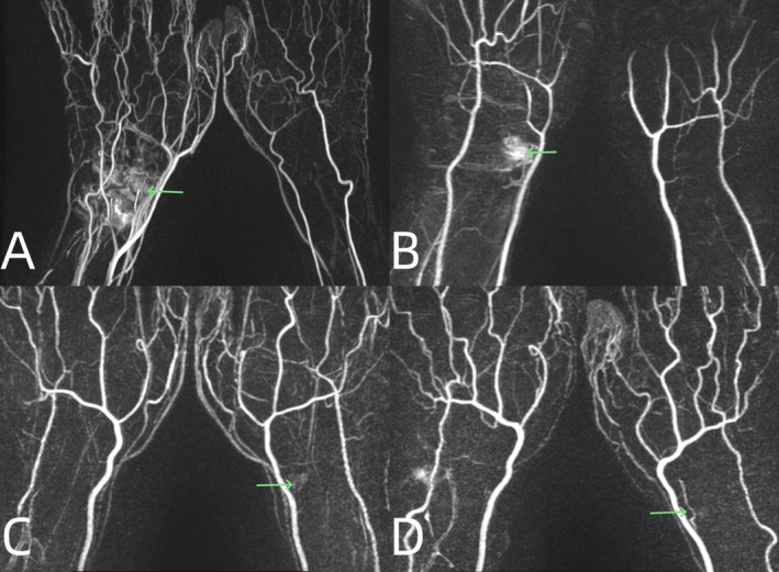
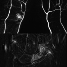
Results: 31 of 32 patients showed contrast media accumulation in the wrist joints on the affected sides. The filling time of contrast media on the affected sides was 5.6 (6.05-1) s quicker than on the healthy sides. The diameters of the radial arteries on the affected side increased by 12.8% (SD, 18.4%) compared to those on the healthy side (p = 0.002). All the patients had visible scaphoid nutrient arteries originating from the radial arteries on the affected side. The number of visible arteries on the healthy side was lower. Blood supply to the scaphoid was not related to the patient's sex, injury side, or fracture site. The increase in blood supply at the proximal fragment in older patients was less than that in young individuals (p = 0.015). Blood supply to the proximal and distal fragments of the scaphoid increased after fracture (p < 0.05). Within 1 month after the fracture, the increase in blood supply at the proximal fragment was less than that at the distal fragment, and it increased significantly after 1 month (p = 0.014). However, long-term nonunion (more than 4 years) leads to a decrease in proximal blood supply.
Conclusions: The unique blood supply pattern of the scaphoid and fracture sites might not be the cause of ischemia after a fracture. This could be due to prolonged nonhealing, which leads to proximal ischemia.
期刊介绍:
Orthopaedic Surgery (OS) is the official journal of the Chinese Orthopaedic Association, focusing on all aspects of orthopaedic technique and surgery.
The journal publishes peer-reviewed articles in the following categories: Original Articles, Clinical Articles, Review Articles, Guidelines, Editorials, Commentaries, Surgical Techniques, Case Reports and Meeting Reports.
 求助内容:
求助内容: 应助结果提醒方式:


