{"title":"Radiographic Anatomy and Clinical Value of the Modified Corner Approach in Interlaminar Endoscopic Lumbar Discectomy.","authors":"Sizheng Zhan, Haoning Ma, Yuming Wang, Ping Yi, Xiangsheng Tang","doi":"10.1111/os.70143","DOIUrl":null,"url":null,"abstract":"<p><strong>Objective: </strong>The first step of interlaminar endoscopic lumbar discectomy (IELD) is puncture localization, which lacks standardized protocols and requires a significant learning curve. To address this, we developed a modified corner approach targeting the junction of the S1 superior endplate and facet joint. This study aims to characterize the radiographic anatomy and assess the clinical utility of this modified approach.</p><p><strong>Method: </strong>Computed tomography (CT) and magnetic resonance imaging (MRI) data from 100 patients were analyzed to measure distances between the target and adjacent structures (dura sac, pedicle, L5 nerve, and S1 nerve). The learning curve of interlaminar endoscopic lumbar discectomy (IELD) surgery based on the modified corner approach was determined by prospectively collecting data from 80 patients.</p><p><strong>Results: </strong>The mean distance between the target and the dura sac was 4.59 ± 1.74 mm. The mean distance between the target and the inferior border of the L5 nerve was 10.14 ± 1.72 mm, rang from 7.52 to 13.54 mm. The mean distance between the target and the outer edge of the S1 nerve was 0.51 ± 0.91 mm, rang from -0.12 to 2 mm. The mean distance between the target and the inner edge of the S1 pedicle was 3.77 ± 1.04 mm. The distance between the target and the dura sac and the inner edge of the pedicle is mainly affected by the patient's age.</p><p><strong>Conclusion: </strong>The modified corner approach is a simple, safe, and repeatable surgical approach with the intersection of the superior endplate and facet joint as the puncture target. For patients without or with mild facet joint degeneration, the puncture target can be appropriately moved inward by 2 mm.</p>","PeriodicalId":19566,"journal":{"name":"Orthopaedic Surgery","volume":" ","pages":"2640-2646"},"PeriodicalIF":2.1000,"publicationDate":"2025-09-01","publicationTypes":"Journal Article","fieldsOfStudy":null,"isOpenAccess":false,"openAccessPdf":"https://www.ncbi.nlm.nih.gov/pmc/articles/PMC12404854/pdf/","citationCount":"0","resultStr":null,"platform":"Semanticscholar","paperid":null,"PeriodicalName":"Orthopaedic Surgery","FirstCategoryId":"3","ListUrlMain":"https://doi.org/10.1111/os.70143","RegionNum":2,"RegionCategory":"医学","ArticlePicture":[],"TitleCN":null,"AbstractTextCN":null,"PMCID":null,"EPubDate":"2025/7/31 0:00:00","PubModel":"Epub","JCR":"Q2","JCRName":"ORTHOPEDICS","Score":null,"Total":0}
引用次数: 0
Abstract
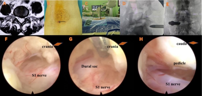
Objective: The first step of interlaminar endoscopic lumbar discectomy (IELD) is puncture localization, which lacks standardized protocols and requires a significant learning curve. To address this, we developed a modified corner approach targeting the junction of the S1 superior endplate and facet joint. This study aims to characterize the radiographic anatomy and assess the clinical utility of this modified approach.
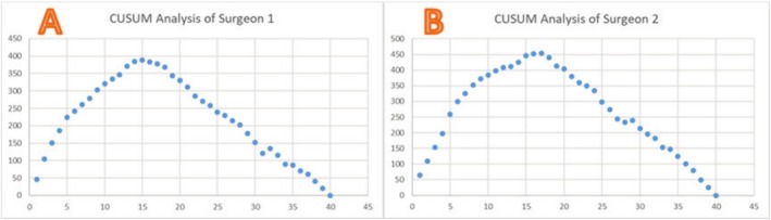
Method: Computed tomography (CT) and magnetic resonance imaging (MRI) data from 100 patients were analyzed to measure distances between the target and adjacent structures (dura sac, pedicle, L5 nerve, and S1 nerve). The learning curve of interlaminar endoscopic lumbar discectomy (IELD) surgery based on the modified corner approach was determined by prospectively collecting data from 80 patients.
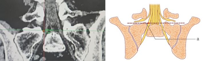
Results: The mean distance between the target and the dura sac was 4.59 ± 1.74 mm. The mean distance between the target and the inferior border of the L5 nerve was 10.14 ± 1.72 mm, rang from 7.52 to 13.54 mm. The mean distance between the target and the outer edge of the S1 nerve was 0.51 ± 0.91 mm, rang from -0.12 to 2 mm. The mean distance between the target and the inner edge of the S1 pedicle was 3.77 ± 1.04 mm. The distance between the target and the dura sac and the inner edge of the pedicle is mainly affected by the patient's age.
Conclusion: The modified corner approach is a simple, safe, and repeatable surgical approach with the intersection of the superior endplate and facet joint as the puncture target. For patients without or with mild facet joint degeneration, the puncture target can be appropriately moved inward by 2 mm.
期刊介绍:
Orthopaedic Surgery (OS) is the official journal of the Chinese Orthopaedic Association, focusing on all aspects of orthopaedic technique and surgery.
The journal publishes peer-reviewed articles in the following categories: Original Articles, Clinical Articles, Review Articles, Guidelines, Editorials, Commentaries, Surgical Techniques, Case Reports and Meeting Reports.
 求助内容:
求助内容: 应助结果提醒方式:


