Haijuan Lv, Yu Zhang, Xinyu Wang, Hu Liu, Hongwei Zhao
{"title":"Correlation Between Hepatic Iron and Fat in Hemodialysis Patients: A Quantitative MRI Analysis.","authors":"Haijuan Lv, Yu Zhang, Xinyu Wang, Hu Liu, Hongwei Zhao","doi":"10.1016/j.xkme.2025.101035","DOIUrl":null,"url":null,"abstract":"<p><strong>Rationale & objective: </strong>Hepatic iron overload and steatosis are common in hemodialysis patients with anemia who are receiving iron therapy. This study aimed to investigate the relationship between hepatic iron overload and steatosis using magnetic resonance imaging (MRI) while also measuring iron content in extrahepatic organs.</p><p><strong>Study design: </strong>Prospective cohort study.</p><p><strong>Setting & participants: </strong>Fifty-three hemodialysis patients with anemia and a history of iron therapy, along with 45 healthy controls, were recruited from a single center between July 2023 and February 2024.</p><p><strong>Interventions: </strong>All participants underwent 3T MRI with mDIXON-Quant sequences to measure R2∗ and proton density fat fraction (PDFF) values in the liver, pancreas, spleen, and vertebral bone marrow.</p><p><strong>Outcomes: </strong>The primary outcome was the correlation between hepatic R2∗ and PDFF. Secondary analyses evaluated the distribution of iron and fat in extrahepatic organs.</p><p><strong>Results: </strong>Severe hepatic iron overload (R2∗ > 70.1 s<sup>-1</sup>) was observed in 52.8% of patients. Among these patients, hepatic R2∗ was strongly correlated with PDFF (<i>r</i> = 0.67, <i>P</i> < 0.001), whereas a weaker correlation was noted in nonsevere cases (<i>r</i> = 0.16, <i>P</i> = 0.43). Serum ferritin levels were highly correlated with R2∗ in the liver (<i>r</i> = 0.87, <i>P</i> < 0.001), pancreas (<i>r</i> = 0.71, <i>P</i> < 0.001), and spleen (<i>r</i> = 0.78, <i>P</i> < 0.001).</p><p><strong>Limitations: </strong>This single-center study included a relatively small sample size, lacked adjustment for potential confounders, and did not include long-term follow-up.</p><p><strong>Conclusions: </strong>Severe hepatic iron overload is closely associated with elevated liver fat content in hemodialysis patients. These MRI findings may inform more personalized iron therapy strategies by enabling a comprehensive assessment of both hepatic and extrahepatic iron deposition, potentially mitigating treatment-related complications.</p>","PeriodicalId":17885,"journal":{"name":"Kidney Medicine","volume":"7 8","pages":"101035"},"PeriodicalIF":3.4000,"publicationDate":"2025-05-20","publicationTypes":"Journal Article","fieldsOfStudy":null,"isOpenAccess":false,"openAccessPdf":"https://www.ncbi.nlm.nih.gov/pmc/articles/PMC12304958/pdf/","citationCount":"0","resultStr":null,"platform":"Semanticscholar","paperid":null,"PeriodicalName":"Kidney Medicine","FirstCategoryId":"1085","ListUrlMain":"https://doi.org/10.1016/j.xkme.2025.101035","RegionNum":0,"RegionCategory":null,"ArticlePicture":[],"TitleCN":null,"AbstractTextCN":null,"PMCID":null,"EPubDate":"2025/8/1 0:00:00","PubModel":"eCollection","JCR":"Q1","JCRName":"UROLOGY & NEPHROLOGY","Score":null,"Total":0}
引用次数: 0
Abstract
Rationale & objective: Hepatic iron overload and steatosis are common in hemodialysis patients with anemia who are receiving iron therapy. This study aimed to investigate the relationship between hepatic iron overload and steatosis using magnetic resonance imaging (MRI) while also measuring iron content in extrahepatic organs.
Study design: Prospective cohort study.
Setting & participants: Fifty-three hemodialysis patients with anemia and a history of iron therapy, along with 45 healthy controls, were recruited from a single center between July 2023 and February 2024.
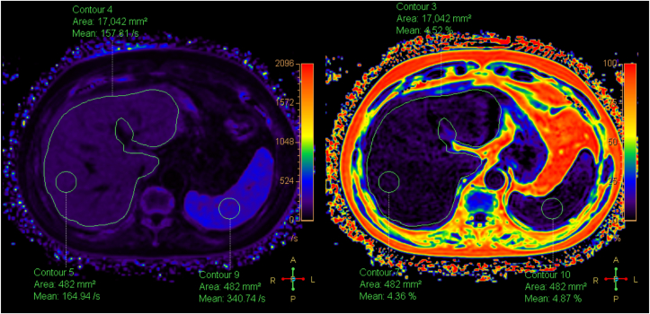
Interventions: All participants underwent 3T MRI with mDIXON-Quant sequences to measure R2∗ and proton density fat fraction (PDFF) values in the liver, pancreas, spleen, and vertebral bone marrow.
Outcomes: The primary outcome was the correlation between hepatic R2∗ and PDFF. Secondary analyses evaluated the distribution of iron and fat in extrahepatic organs.
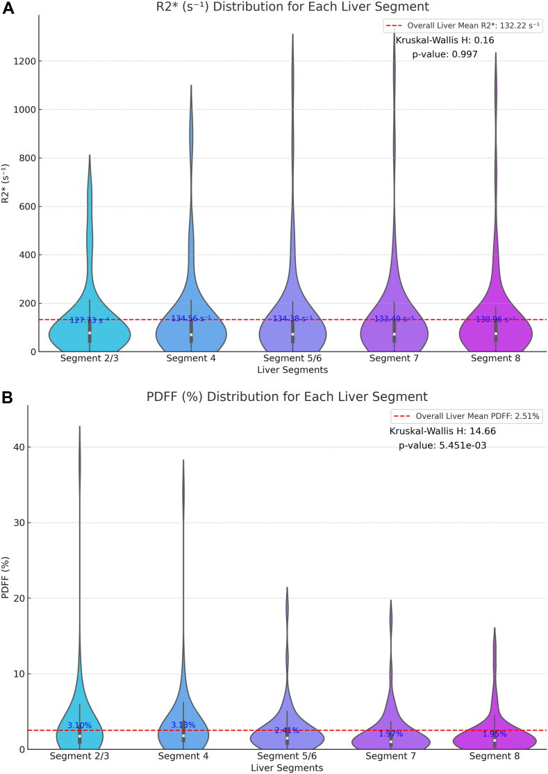
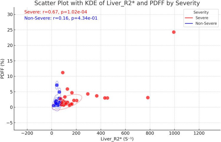
Results: Severe hepatic iron overload (R2∗ > 70.1 s-1) was observed in 52.8% of patients. Among these patients, hepatic R2∗ was strongly correlated with PDFF (r = 0.67, P < 0.001), whereas a weaker correlation was noted in nonsevere cases (r = 0.16, P = 0.43). Serum ferritin levels were highly correlated with R2∗ in the liver (r = 0.87, P < 0.001), pancreas (r = 0.71, P < 0.001), and spleen (r = 0.78, P < 0.001).
Limitations: This single-center study included a relatively small sample size, lacked adjustment for potential confounders, and did not include long-term follow-up.
Conclusions: Severe hepatic iron overload is closely associated with elevated liver fat content in hemodialysis patients. These MRI findings may inform more personalized iron therapy strategies by enabling a comprehensive assessment of both hepatic and extrahepatic iron deposition, potentially mitigating treatment-related complications.
 求助内容:
求助内容: 应助结果提醒方式:


