Zhixin Sun, Jacqueline M Godbe, Alexander Zheleznyak, Brad Manion, Junhao Hu, Julie L Prior, Kathleen Duncan, Ulugbek S Kamilov, Monica Shokeen
{"title":"Advanced quantification pipeline reveals new spatial and temporal tumor characteristics in preclinical multiple myeloma.","authors":"Zhixin Sun, Jacqueline M Godbe, Alexander Zheleznyak, Brad Manion, Junhao Hu, Julie L Prior, Kathleen Duncan, Ulugbek S Kamilov, Monica Shokeen","doi":"10.1186/s13550-025-01291-x","DOIUrl":null,"url":null,"abstract":"<p><strong>Background: </strong>Radiological imaging plays an indispensable role in both preclinical and clinical studies of multiple myeloma (MM). However, manual quantification in longitudinal small animal PET/CT is limited by annotator bias, signal artifacts from urinary/fecal excretion, and voxel misalignment due to non-rigid registration. To address these challenges and improve characterization of tumor biology, we developed a semi-automated PET/CT quantification pipeline targeting defined regions of interest (ROIs) within the bone marrow-rich mouse skeleton, achieving sub-organ spatial resolution, including in anatomically complex sites such as the pelvis. We applied this MM-specific preclinical pipeline to analyze tumor distribution in a longitudinal molecular PET study using an immunocompetent mouse model of skeletally disseminated MM. An Attention U-Net was trained to segment the thoracolumbar spine, pelvis and pelvic joints, sacrum, and femurs from 2D CT slices. A custom algorithm masked spillover signal from physiological excretion, and a PCA-based projection was used to map tumor distribution along the skeletal axis. Quantification metrics included mean and maximum standardized uptake values (SUV<sub>mean</sub>, SUV<sub>max</sub>) from PET and Hounsfield Units (HU) from CT to assess tumor burden, spatiotemporal tumor distribution, and bone involvement.</p><p><strong>Results: </strong>Tumor burden localized preferentially to skeletal regions near joints. Using precise CT-based alignment (DICE = 0.966 ± 0.005), we detected early disease progression and aggressive phenotypes. A marked increase in tumor uptake was observed by day 18 post-implantation, with significant SUV<sub>mean</sub> increases in the spine (p = 0.012), left/right femurs (p = 0.007/0.006), pelvis and pelvic joints (p = 0.018), and sacrum (p = 0.02). Notably, sex-based differences were identified: female mice showed greater bone loss near the hip joint at later stages, with significant HU<sub>mean</sub> reductions at days 25 (p = 0.008) and 32 (p = 0.002).</p><p><strong>Conclusions: </strong>This pipeline enables reproducible, anatomically precise quantification of region-specific trends in MM progression, including joint-specific lesion tropism and sex-based differences, from longitudinal PET/CT scans. By mitigating common challenges such as excretion artifacts and inconsistent mouse positioning, our approach overcomes limitations of manual analysis and enhances evaluation of tumor biology and treatment response in preclinical models of bone-involved cancers.</p>","PeriodicalId":11611,"journal":{"name":"EJNMMI Research","volume":"15 1","pages":"95"},"PeriodicalIF":3.1000,"publicationDate":"2025-07-31","publicationTypes":"Journal Article","fieldsOfStudy":null,"isOpenAccess":false,"openAccessPdf":"https://www.ncbi.nlm.nih.gov/pmc/articles/PMC12314142/pdf/","citationCount":"0","resultStr":null,"platform":"Semanticscholar","paperid":null,"PeriodicalName":"EJNMMI Research","FirstCategoryId":"3","ListUrlMain":"https://doi.org/10.1186/s13550-025-01291-x","RegionNum":3,"RegionCategory":"医学","ArticlePicture":[],"TitleCN":null,"AbstractTextCN":null,"PMCID":null,"EPubDate":"","PubModel":"","JCR":"Q1","JCRName":"RADIOLOGY, NUCLEAR MEDICINE & MEDICAL IMAGING","Score":null,"Total":0}
引用次数: 0
Abstract
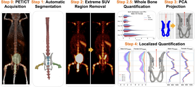
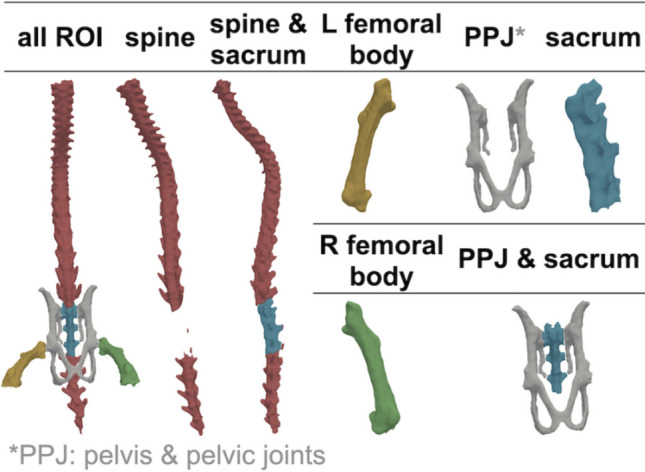
Background: Radiological imaging plays an indispensable role in both preclinical and clinical studies of multiple myeloma (MM). However, manual quantification in longitudinal small animal PET/CT is limited by annotator bias, signal artifacts from urinary/fecal excretion, and voxel misalignment due to non-rigid registration. To address these challenges and improve characterization of tumor biology, we developed a semi-automated PET/CT quantification pipeline targeting defined regions of interest (ROIs) within the bone marrow-rich mouse skeleton, achieving sub-organ spatial resolution, including in anatomically complex sites such as the pelvis. We applied this MM-specific preclinical pipeline to analyze tumor distribution in a longitudinal molecular PET study using an immunocompetent mouse model of skeletally disseminated MM. An Attention U-Net was trained to segment the thoracolumbar spine, pelvis and pelvic joints, sacrum, and femurs from 2D CT slices. A custom algorithm masked spillover signal from physiological excretion, and a PCA-based projection was used to map tumor distribution along the skeletal axis. Quantification metrics included mean and maximum standardized uptake values (SUVmean, SUVmax) from PET and Hounsfield Units (HU) from CT to assess tumor burden, spatiotemporal tumor distribution, and bone involvement.
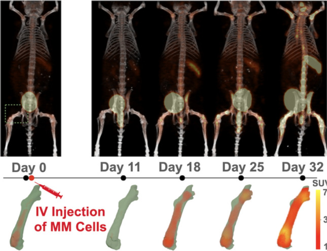
Results: Tumor burden localized preferentially to skeletal regions near joints. Using precise CT-based alignment (DICE = 0.966 ± 0.005), we detected early disease progression and aggressive phenotypes. A marked increase in tumor uptake was observed by day 18 post-implantation, with significant SUVmean increases in the spine (p = 0.012), left/right femurs (p = 0.007/0.006), pelvis and pelvic joints (p = 0.018), and sacrum (p = 0.02). Notably, sex-based differences were identified: female mice showed greater bone loss near the hip joint at later stages, with significant HUmean reductions at days 25 (p = 0.008) and 32 (p = 0.002).
Conclusions: This pipeline enables reproducible, anatomically precise quantification of region-specific trends in MM progression, including joint-specific lesion tropism and sex-based differences, from longitudinal PET/CT scans. By mitigating common challenges such as excretion artifacts and inconsistent mouse positioning, our approach overcomes limitations of manual analysis and enhances evaluation of tumor biology and treatment response in preclinical models of bone-involved cancers.
EJNMMI ResearchRADIOLOGY, NUCLEAR MEDICINE & MEDICAL IMAGING&nb-
CiteScore
5.90
自引率
3.10%
发文量
72
审稿时长
13 weeks
期刊介绍:
EJNMMI Research publishes new basic, translational and clinical research in the field of nuclear medicine and molecular imaging. Regular features include original research articles, rapid communication of preliminary data on innovative research, interesting case reports, editorials, and letters to the editor. Educational articles on basic sciences, fundamental aspects and controversy related to pre-clinical and clinical research or ethical aspects of research are also welcome. Timely reviews provide updates on current applications, issues in imaging research and translational aspects of nuclear medicine and molecular imaging technologies.
The main emphasis is placed on the development of targeted imaging with radiopharmaceuticals within the broader context of molecular probes to enhance understanding and characterisation of the complex biological processes underlying disease and to develop, test and guide new treatment modalities, including radionuclide therapy.

 求助内容:
求助内容: 应助结果提醒方式:


