Lucía Sanabria Greciano, Ana Fernández Alfonso, Begoña Peinado Iribar, Raquel Cano Alonso, Ana Álvarez Vázquez, Vicente Martínez de Vega Fernández
{"title":"Accessory spleen torsion: a hidden etiology of acute abdominal emergency.","authors":"Lucía Sanabria Greciano, Ana Fernández Alfonso, Begoña Peinado Iribar, Raquel Cano Alonso, Ana Álvarez Vázquez, Vicente Martínez de Vega Fernández","doi":"10.1093/bjrcr/uaaf035","DOIUrl":null,"url":null,"abstract":"<p><p>Accessory spleen torsion is a rare but important cause of acute abdominal pain, often presenting with non-specific symptoms that overlap with more common abdominal pathologies. This case report discusses a 19-year-old female who presented with left-sided flank pain and leucocytosis. Imaging with abdominal CT and MRI revealed a well-defined lesion near the spleen and kidney, with mild vascular engorgement and surrounding inflammation. While these findings raised suspicion for accessory spleen torsion, the diagnosis was not immediately clear. The lesion's location, vascular congestion, and absence of typical characteristics for other pathologies, such as haematomas, abscesses, mesothelial cysts, or lymphangiomas pointed towards torsion, but confirmation required surgical intervention. During laparoscopic exploration, a 5 cm accessory spleen with ischaemic changes due to torsion of its pedicle was identified and successfully removed without complications. Accessory spleens, present in 10%-30% of the population, are usually asymptomatic but can become problematic if torsion, rupture, or infarction occurs. Imaging plays a critical role in identifying torsion, with CT and MRI revealing the characteristic \"whirlpool sign\" and vascular congestion. Early recognition is crucial to prevent complications such as necrosis and rupture, and surgical intervention, typically laparoscopic splenectomy, is the treatment of choice. This case highlights the importance of considering accessory spleen torsion in the differential diagnosis of acute abdominal pain, particularly in young patients with non-specific symptoms. Awareness of this condition can improve early diagnosis and outcomes, preventing severe consequences.</p>","PeriodicalId":45216,"journal":{"name":"BJR Case Reports","volume":"11 4","pages":"uaaf035"},"PeriodicalIF":0.5000,"publicationDate":"2025-07-16","publicationTypes":"Journal Article","fieldsOfStudy":null,"isOpenAccess":false,"openAccessPdf":"https://www.ncbi.nlm.nih.gov/pmc/articles/PMC12303862/pdf/","citationCount":"0","resultStr":null,"platform":"Semanticscholar","paperid":null,"PeriodicalName":"BJR Case Reports","FirstCategoryId":"1085","ListUrlMain":"https://doi.org/10.1093/bjrcr/uaaf035","RegionNum":0,"RegionCategory":null,"ArticlePicture":[],"TitleCN":null,"AbstractTextCN":null,"PMCID":null,"EPubDate":"2025/7/1 0:00:00","PubModel":"eCollection","JCR":"Q4","JCRName":"RADIOLOGY, NUCLEAR MEDICINE & MEDICAL IMAGING","Score":null,"Total":0}
引用次数: 0
Abstract
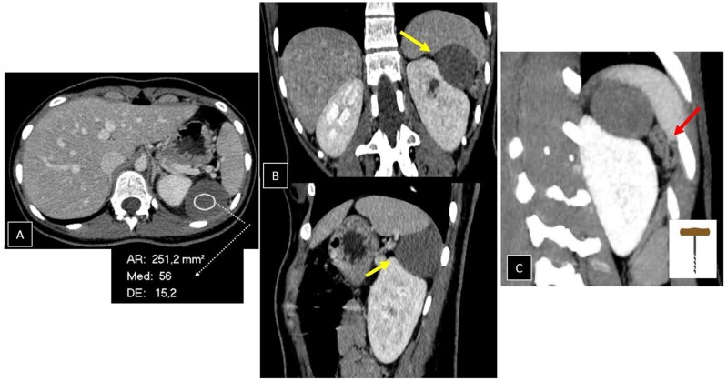
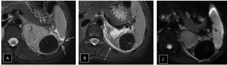
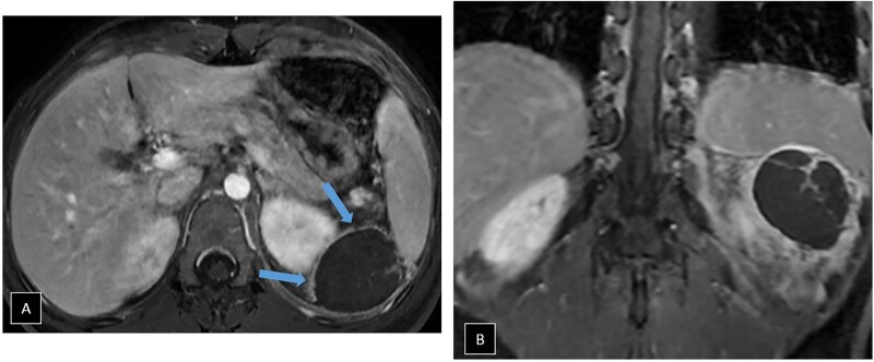
Accessory spleen torsion is a rare but important cause of acute abdominal pain, often presenting with non-specific symptoms that overlap with more common abdominal pathologies. This case report discusses a 19-year-old female who presented with left-sided flank pain and leucocytosis. Imaging with abdominal CT and MRI revealed a well-defined lesion near the spleen and kidney, with mild vascular engorgement and surrounding inflammation. While these findings raised suspicion for accessory spleen torsion, the diagnosis was not immediately clear. The lesion's location, vascular congestion, and absence of typical characteristics for other pathologies, such as haematomas, abscesses, mesothelial cysts, or lymphangiomas pointed towards torsion, but confirmation required surgical intervention. During laparoscopic exploration, a 5 cm accessory spleen with ischaemic changes due to torsion of its pedicle was identified and successfully removed without complications. Accessory spleens, present in 10%-30% of the population, are usually asymptomatic but can become problematic if torsion, rupture, or infarction occurs. Imaging plays a critical role in identifying torsion, with CT and MRI revealing the characteristic "whirlpool sign" and vascular congestion. Early recognition is crucial to prevent complications such as necrosis and rupture, and surgical intervention, typically laparoscopic splenectomy, is the treatment of choice. This case highlights the importance of considering accessory spleen torsion in the differential diagnosis of acute abdominal pain, particularly in young patients with non-specific symptoms. Awareness of this condition can improve early diagnosis and outcomes, preventing severe consequences.

 求助内容:
求助内容: 应助结果提醒方式:


