{"title":"Distinct Roles of HHLA2 and PD-L1 in the Immune Cell and Prognosis of Hepatocellular Carcinoma.","authors":"Chun-Hua Wang, Shi-Lu Chen, Xia Yang, Ting Wu, Li-Li Liu, Jing-Ping Yun","doi":"10.2147/JHC.S513033","DOIUrl":null,"url":null,"abstract":"<p><strong>Background: </strong>HHLA2, a member of the B7 family, is extensively expressed in various cancers and plays a pivotal role in modulating the immune microenvironment. However, its prognostic significance in hepatocellular carcinoma (HCC) remains poorly understood. This study aims to elucidate the expression patterns of HHLA2 and PD-L1 in HCC, their associations with tumor-infiltrating lymphocytes (TILs), and their impact on clinical outcomes.</p><p><strong>Methods: </strong>Immunohistochemistry (IHC) was employed to evaluate HHLA2 and PD-L1 expression in 547 HCC tissue samples. PD-L1 positivity was defined as ≥1% membranous or cytoplasmic staining. Hematoxylin and eosin (H&E) staining was utilized to quantify TILs (percentage/area), while IHC was used to measure the densities of CD3+, CD4+, and CD8+ TILs (cells/mm²).</p><p><strong>Results: </strong>HHLA2 and PD-L1 exhibited similar positivity rates. HHLA2 positivity was associated with older age, lower alpha-fetoprotein (AFP) levels, well-differentiated tumors, and improved overall survival (OS). HHLA2 expression was inversely correlated with stromal TIL density. In contrast, tumor cell (TC)-PD-L1 and inflammatory cell (IC)-PD-L1 positivity were positively correlated with higher stromal TIL density and increased levels of CD3+, CD4+, and CD8+ TILs. Patients with HHLA2(+)/PD-L1(-) status demonstrated the longest OS. A novel classification system based on HHLA2/PD-L1 expression identified distinct immune profiles and prognostic subgroups.</p><p><strong>Conclusion: </strong>HHLA2 significantly influences the immune microenvironment of HCC and serves as an independent prognostic marker. The combined assessment of HHLA2 and PD-L1 expression facilitates risk stratification, providing a framework to optimize immunotherapy strategies. These findings contribute to the advancement of precision medicine in the management of HCC.</p>","PeriodicalId":15906,"journal":{"name":"Journal of Hepatocellular Carcinoma","volume":"12 ","pages":"1633-1645"},"PeriodicalIF":3.4000,"publicationDate":"2025-07-25","publicationTypes":"Journal Article","fieldsOfStudy":null,"isOpenAccess":false,"openAccessPdf":"https://www.ncbi.nlm.nih.gov/pmc/articles/PMC12306544/pdf/","citationCount":"0","resultStr":null,"platform":"Semanticscholar","paperid":null,"PeriodicalName":"Journal of Hepatocellular Carcinoma","FirstCategoryId":"3","ListUrlMain":"https://doi.org/10.2147/JHC.S513033","RegionNum":3,"RegionCategory":"医学","ArticlePicture":[],"TitleCN":null,"AbstractTextCN":null,"PMCID":null,"EPubDate":"2025/1/1 0:00:00","PubModel":"eCollection","JCR":"Q2","JCRName":"ONCOLOGY","Score":null,"Total":0}
引用次数: 0
Abstract
Background: HHLA2, a member of the B7 family, is extensively expressed in various cancers and plays a pivotal role in modulating the immune microenvironment. However, its prognostic significance in hepatocellular carcinoma (HCC) remains poorly understood. This study aims to elucidate the expression patterns of HHLA2 and PD-L1 in HCC, their associations with tumor-infiltrating lymphocytes (TILs), and their impact on clinical outcomes.
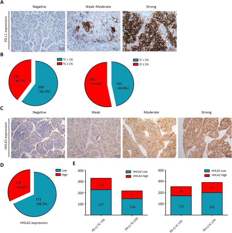
Methods: Immunohistochemistry (IHC) was employed to evaluate HHLA2 and PD-L1 expression in 547 HCC tissue samples. PD-L1 positivity was defined as ≥1% membranous or cytoplasmic staining. Hematoxylin and eosin (H&E) staining was utilized to quantify TILs (percentage/area), while IHC was used to measure the densities of CD3+, CD4+, and CD8+ TILs (cells/mm²).
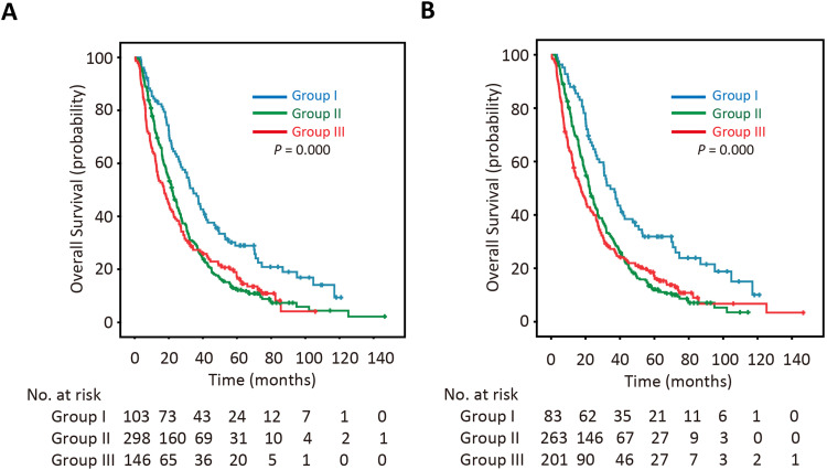
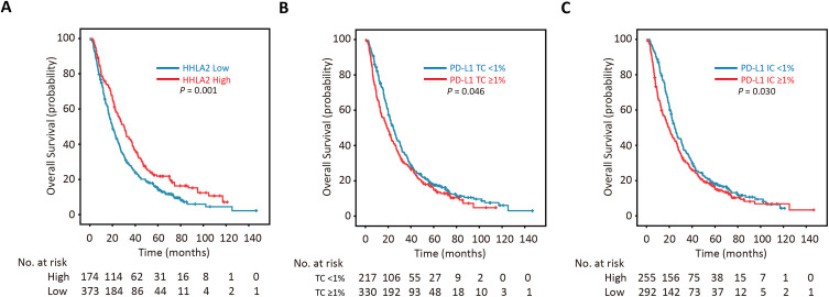
Results: HHLA2 and PD-L1 exhibited similar positivity rates. HHLA2 positivity was associated with older age, lower alpha-fetoprotein (AFP) levels, well-differentiated tumors, and improved overall survival (OS). HHLA2 expression was inversely correlated with stromal TIL density. In contrast, tumor cell (TC)-PD-L1 and inflammatory cell (IC)-PD-L1 positivity were positively correlated with higher stromal TIL density and increased levels of CD3+, CD4+, and CD8+ TILs. Patients with HHLA2(+)/PD-L1(-) status demonstrated the longest OS. A novel classification system based on HHLA2/PD-L1 expression identified distinct immune profiles and prognostic subgroups.
Conclusion: HHLA2 significantly influences the immune microenvironment of HCC and serves as an independent prognostic marker. The combined assessment of HHLA2 and PD-L1 expression facilitates risk stratification, providing a framework to optimize immunotherapy strategies. These findings contribute to the advancement of precision medicine in the management of HCC.
 求助内容:
求助内容: 应助结果提醒方式:


