Sheng-Chieh Chiu, Jose Angelo U Perucho, Yu-Hua Dean Fang
{"title":"Motion correction of simultaneous brain PET/MR images based on tracer uptake characteristics.","authors":"Sheng-Chieh Chiu, Jose Angelo U Perucho, Yu-Hua Dean Fang","doi":"10.1186/s40658-025-00789-6","DOIUrl":null,"url":null,"abstract":"<p><strong>Background: </strong>Simultaneous PET/MR imaging enables precise anatomical localization and PET quantification by reducing PET-to-MR misalignments. However, involuntary motion during scans may still cause misalignment and quantification imprecision. Current mutual information (MI)-based co-registration methods do not account for the tissue-specific uptake patterns of PET and therefore could result in suboptimal alignment. To address this, we proposed a novel image co-registration method, namely the tracer characteristic-based co-registration (TCBC) method, which takes advantage of specific PET uptake patterns within a selected anatomical region to improve the image alignment and PET quantification.</p><p><strong>Results: </strong>TCBC was evaluated using simulation and in vivo <sup>18</sup>F-Florbetapir PET/MR data from the OASIS-3 dataset. In simulations, TCBC demonstrated superior alignment accuracy with lower root mean square error and higher R-squared values compared to the conventional MI-based co-registration from FreeSurfer in recovering the simulated patient motion. In the retrospective human study, we evaluated the detectability of age-related amyloid burden in healthy controls under different co-registration methods as a demonstrative use case. TCBC significantly enhanced the detectability of age-related amyloid burden with stronger correlations across all five regions of evaluation, such as the medial orbitofrontal cortex (p < 0.001), precuneus (p = 0.004), and early amyloid-β composite (p = 0.002), compared to FSMC (p = 0.004, 0.007, and 0.006, respectively) and uncorrected (p = 0.378, 0.023, and 0.039, respectively) methods. Bootstrap analyses also confirmed TCBC's robustness in smaller samples, yielding tighter confidence intervals and lower means of p-values, such as 0.032 (95% CI: 0.029-0.035) in the precuneus and 0.008 (CI: 0.007-0.010) in the medial orbitofrontal cortex, outperforming FSMC (p = 0.046 with CI: 0.042-0.049, and p = 0.040 with CI: 0.036-0.044, respectively).</p><p><strong>Conclusions: </strong>The TCBC method reduces image misalignment, improves PET quantification, and may have a good potential for being applied to both research and clinical studies with simultaneous brain PET/MR.</p><p><strong>Clinical trial number: </strong>Not applicable.</p>","PeriodicalId":11559,"journal":{"name":"EJNMMI Physics","volume":"12 1","pages":"75"},"PeriodicalIF":3.2000,"publicationDate":"2025-07-30","publicationTypes":"Journal Article","fieldsOfStudy":null,"isOpenAccess":false,"openAccessPdf":"https://www.ncbi.nlm.nih.gov/pmc/articles/PMC12311070/pdf/","citationCount":"0","resultStr":null,"platform":"Semanticscholar","paperid":null,"PeriodicalName":"EJNMMI Physics","FirstCategoryId":"3","ListUrlMain":"https://doi.org/10.1186/s40658-025-00789-6","RegionNum":2,"RegionCategory":"医学","ArticlePicture":[],"TitleCN":null,"AbstractTextCN":null,"PMCID":null,"EPubDate":"","PubModel":"","JCR":"Q2","JCRName":"RADIOLOGY, NUCLEAR MEDICINE & MEDICAL IMAGING","Score":null,"Total":0}
引用次数: 0
Abstract
Background: Simultaneous PET/MR imaging enables precise anatomical localization and PET quantification by reducing PET-to-MR misalignments. However, involuntary motion during scans may still cause misalignment and quantification imprecision. Current mutual information (MI)-based co-registration methods do not account for the tissue-specific uptake patterns of PET and therefore could result in suboptimal alignment. To address this, we proposed a novel image co-registration method, namely the tracer characteristic-based co-registration (TCBC) method, which takes advantage of specific PET uptake patterns within a selected anatomical region to improve the image alignment and PET quantification.
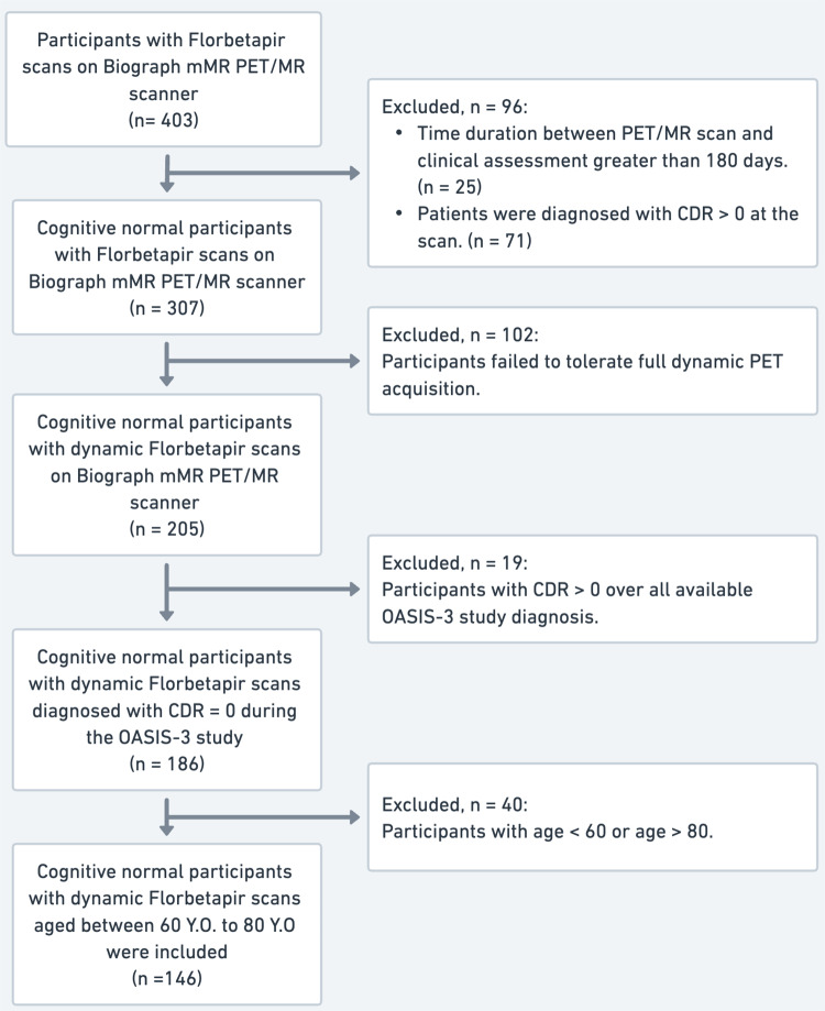
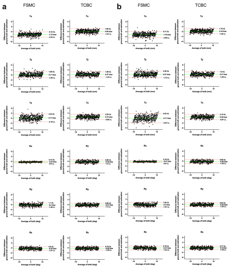
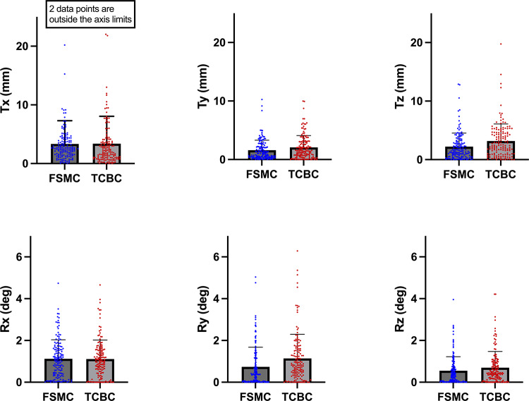
Results: TCBC was evaluated using simulation and in vivo 18F-Florbetapir PET/MR data from the OASIS-3 dataset. In simulations, TCBC demonstrated superior alignment accuracy with lower root mean square error and higher R-squared values compared to the conventional MI-based co-registration from FreeSurfer in recovering the simulated patient motion. In the retrospective human study, we evaluated the detectability of age-related amyloid burden in healthy controls under different co-registration methods as a demonstrative use case. TCBC significantly enhanced the detectability of age-related amyloid burden with stronger correlations across all five regions of evaluation, such as the medial orbitofrontal cortex (p < 0.001), precuneus (p = 0.004), and early amyloid-β composite (p = 0.002), compared to FSMC (p = 0.004, 0.007, and 0.006, respectively) and uncorrected (p = 0.378, 0.023, and 0.039, respectively) methods. Bootstrap analyses also confirmed TCBC's robustness in smaller samples, yielding tighter confidence intervals and lower means of p-values, such as 0.032 (95% CI: 0.029-0.035) in the precuneus and 0.008 (CI: 0.007-0.010) in the medial orbitofrontal cortex, outperforming FSMC (p = 0.046 with CI: 0.042-0.049, and p = 0.040 with CI: 0.036-0.044, respectively).
Conclusions: The TCBC method reduces image misalignment, improves PET quantification, and may have a good potential for being applied to both research and clinical studies with simultaneous brain PET/MR.
期刊介绍:
EJNMMI Physics is an international platform for scientists, users and adopters of nuclear medicine with a particular interest in physics matters. As a companion journal to the European Journal of Nuclear Medicine and Molecular Imaging, this journal has a multi-disciplinary approach and welcomes original materials and studies with a focus on applied physics and mathematics as well as imaging systems engineering and prototyping in nuclear medicine. This includes physics-driven approaches or algorithms supported by physics that foster early clinical adoption of nuclear medicine imaging and therapy.

 求助内容:
求助内容: 应助结果提醒方式:


