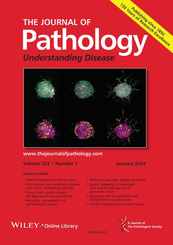
Pyung Hun Park, Lauren E Israel, Michael H Alexander, Grace Tartaglia, Harvey G South, Suhao Han, Joseph M Curry, Adam J Luginbuhl, Andrew P South
下载PDF
{"title":"Partial-EMT cell state correlates with single cell pattern of invasion in head and neck SCC keratinocytes","authors":"Pyung Hun Park, Lauren E Israel, Michael H Alexander, Grace Tartaglia, Harvey G South, Suhao Han, Joseph M Curry, Adam J Luginbuhl, Andrew P South","doi":"10.1002/path.6454","DOIUrl":null,"url":null,"abstract":"<p>The presence of a single metastatic lesion significantly decreases overall survival in patients with head and neck squamous cell carcinoma (HNSCC), and invasion of malignant keratinocytes is one of the initial steps required for HNSCC metastasis. Histological grading of tumor cell invasion predicts outcome in HNSCC, yet the molecular factors that determine the extent of invasion, and subsequent grading are not fully understood. Using a 3D organ culture model and multiple patient-derived HNSCC keratinocytes representing all major anatomical subsites of the disease, we identified a range of cell states that represent a continuum of epithelial-to-mesenchymal (EMT) characteristics. We also demonstrated how these cell states change in response to TGF-beta stimulation and co-culture with cancer-associated fibroblasts in organ cultures. Using 3D culture models that recapitulate the pattern of invasion seen in primary tumors from which the keratinocytes were derived, we identified distinct clusters of partial-EMT marker expression in individual patient HNSCC keratinocyte populations. Partial-EMT transcription factors were correlated with separate invasive characteristics, and we demonstrated that ZEB2 (a known EMT driver) and HIC1 (a novel EMT driver) are central nodes in HNSCC keratinocyte invasion. Collectively, our findings refine the concepts of partial-EMT and tumor cell invasion, and identify potential therapeutic targets for future development. © 2025 The Author(s). <i>The Journal of Pathology</i> published by John Wiley & Sons Ltd on behalf of The Pathological Society of Great Britain and Ireland.</p>","PeriodicalId":232,"journal":{"name":"The Journal of Pathology","volume":"267 1","pages":"92-104"},"PeriodicalIF":5.2000,"publicationDate":"2025-07-30","publicationTypes":"Journal Article","fieldsOfStudy":null,"isOpenAccess":false,"openAccessPdf":"https://www.ncbi.nlm.nih.gov/pmc/articles/PMC12313228/pdf/","citationCount":"0","resultStr":null,"platform":"Semanticscholar","paperid":null,"PeriodicalName":"The Journal of Pathology","FirstCategoryId":"3","ListUrlMain":"https://pathsocjournals.onlinelibrary.wiley.com/doi/10.1002/path.6454","RegionNum":2,"RegionCategory":"医学","ArticlePicture":[],"TitleCN":null,"AbstractTextCN":null,"PMCID":null,"EPubDate":"","PubModel":"","JCR":"Q1","JCRName":"ONCOLOGY","Score":null,"Total":0}
引用次数: 0
引用
批量引用
Abstract
The presence of a single metastatic lesion significantly decreases overall survival in patients with head and neck squamous cell carcinoma (HNSCC), and invasion of malignant keratinocytes is one of the initial steps required for HNSCC metastasis. Histological grading of tumor cell invasion predicts outcome in HNSCC, yet the molecular factors that determine the extent of invasion, and subsequent grading are not fully understood. Using a 3D organ culture model and multiple patient-derived HNSCC keratinocytes representing all major anatomical subsites of the disease, we identified a range of cell states that represent a continuum of epithelial-to-mesenchymal (EMT) characteristics. We also demonstrated how these cell states change in response to TGF-beta stimulation and co-culture with cancer-associated fibroblasts in organ cultures. Using 3D culture models that recapitulate the pattern of invasion seen in primary tumors from which the keratinocytes were derived, we identified distinct clusters of partial-EMT marker expression in individual patient HNSCC keratinocyte populations. Partial-EMT transcription factors were correlated with separate invasive characteristics, and we demonstrated that ZEB2 (a known EMT driver) and HIC1 (a novel EMT driver) are central nodes in HNSCC keratinocyte invasion. Collectively, our findings refine the concepts of partial-EMT and tumor cell invasion, and identify potential therapeutic targets for future development. © 2025 The Author(s). The Journal of Pathology published by John Wiley & Sons Ltd on behalf of The Pathological Society of Great Britain and Ireland.
部分emt细胞状态与头颈部鳞状细胞角质形成细胞侵袭的单细胞模式相关。
单一转移灶的存在显著降低了头颈部鳞状细胞癌(HNSCC)患者的总生存率,恶性角化细胞的侵袭是HNSCC转移的初始步骤之一。肿瘤细胞侵袭的组织学分级预测HNSCC的预后,但决定侵袭程度的分子因素以及随后的分级尚不完全清楚。利用3D器官培养模型和代表该疾病所有主要解剖亚位的多个患者源性HNSCC角化细胞,我们确定了一系列代表上皮-间质(EMT)特征连续体的细胞状态。我们还展示了这些细胞状态如何在器官培养中响应tgf - β刺激和与癌症相关成纤维细胞共培养而发生变化。利用3D培养模型,概括了角质形成细胞来源于原发肿瘤的侵袭模式,我们在单个HNSCC患者角质形成细胞群体中发现了不同的部分emt标记表达簇。部分EMT转录因子与不同的侵袭特征相关,我们证明了ZEB2(一种已知的EMT驱动因子)和HIC1(一种新型的EMT驱动因子)是HNSCC角化细胞侵袭的中心节点。总的来说,我们的发现完善了部分emt和肿瘤细胞侵袭的概念,并确定了未来发展的潜在治疗靶点。©2025作者。《病理学杂志》由John Wiley & Sons Ltd代表大不列颠和爱尔兰病理学会出版。
本文章由计算机程序翻译,如有差异,请以英文原文为准。


 求助内容:
求助内容: 应助结果提醒方式:


