Accelerating cardiac radial-MRI: Fully polar based technique using compressed sensing and deep learning
IF 11.8
1区 医学
Q1 COMPUTER SCIENCE, ARTIFICIAL INTELLIGENCE
引用次数: 0
Abstract
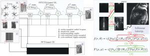
Fast radial-MRI approaches based on compressed sensing (CS) and deep learning (DL) often use non-uniform fast Fourier transform (NUFFT) as the forward imaging operator, which might introduce interpolation errors and reduce image quality. Using the polar Fourier transform (PFT), we developed fully polar CS and DL algorithms for fast 2D cardiac radial-MRI. Our methods directly reconstruct images in polar spatial space from polar k-space data, eliminating frequency interpolation and ensuring an easy-to-compute data consistency term for the DL framework via the variable splitting (VS) scheme. Furthermore, PFT reconstruction produces initial images with fewer artifacts in a reduced field of view, making it a better starting point for CS and DL algorithms, especially for dynamic imaging, where information from a small region of interest is critical, as opposed to NUFFT, which often results in global streaking artifacts. In the cardiac region, PFT-based CS technique outperformed NUFFT-based CS at acceleration rates of 5x (mean SSIM: 0.8831 vs. 0.8526), 10x (0.8195 vs. 0.7981), and 15x (0.7720 vs. 0.7503). Our PFT(VS)-DL technique outperformed the NUFFT(GD)-based DL method, which used unrolled gradient descent with the NUFFT as the forward imaging operator, with mean SSIM scores of 0.8914 versus 0.8617 at 10x and 0.8470 versus 0.8301 at 15x. Radiological assessments revealed that PFT(VS)-based DL scored and at 5x and 10x, whereas NUFFT(GD)-based DL scored and , respectively. Our methods suggest a promising alternative to NUFFT-based fast radial-MRI for dynamic imaging, prioritizing reconstruction quality in a small region of interest over whole image quality.
加速心脏放射mri:使用压缩感知和深度学习的全极性技术
基于压缩感知(CS)和深度学习(DL)的快速径向核磁共振成像方法通常使用非均匀快速傅里叶变换(NUFFT)作为正演成像算子,这可能会引入插值误差,降低图像质量。利用极性傅里叶变换(PFT),我们开发了用于快速二维心脏径向mri的全极性CS和DL算法。我们的方法直接从极坐标k空间数据重建极坐标空间中的图像,消除了频率插值,并通过变量分裂(VS)方案确保了DL框架易于计算的数据一致性项。此外,PFT重建产生的初始图像在缩小的视场中具有更少的伪影,使其成为CS和DL算法的更好起点,特别是对于动态成像,其中来自小区域的信息至关重要,而不是NUFFT,这通常会导致全局条纹伪影。在心脏区域,基于pft的CS技术在加速率上优于基于nufft的CS,分别为5倍(平均SSIM: 0.8831 vs. 0.8526)、10倍(0.8195 vs. 0.7981)和15倍(0.7720 vs. 0.7503)。我们的PFT(VS)-DL技术优于基于NUFFT(GD)的DL方法,该方法使用展开梯度下降和NUFFT作为正演成像算子,平均SSIM分数为0.8914比0.8617在10倍和0.8470比0.8301在15倍。放射学评估显示,基于PFT(VS)的DL在5倍和10倍时分别为2.9±0.30和2.73±0.45,而基于NUFFT(GD)的DL分别为2.7±0.47和2.40±0.50。我们的方法为动态成像提供了一种有希望的替代基于nufft的快速径向mri,优先考虑小区域的重建质量而不是整个图像质量。
本文章由计算机程序翻译,如有差异,请以英文原文为准。
求助全文
约1分钟内获得全文
求助全文
来源期刊

Medical image analysis
工程技术-工程:生物医学
CiteScore
22.10
自引率
6.40%
发文量
309
审稿时长
6.6 months
期刊介绍:
Medical Image Analysis serves as a platform for sharing new research findings in the realm of medical and biological image analysis, with a focus on applications of computer vision, virtual reality, and robotics to biomedical imaging challenges. The journal prioritizes the publication of high-quality, original papers contributing to the fundamental science of processing, analyzing, and utilizing medical and biological images. It welcomes approaches utilizing biomedical image datasets across all spatial scales, from molecular/cellular imaging to tissue/organ imaging.
 求助内容:
求助内容: 应助结果提醒方式:


