Somayeh Meysami, Saurabh Garg, Sam Hashemi, Nasrin Akbari, Ahmed Gouda, Yosef Gavriel Chodakiewitz, Thanh Duc Nguyen, Rajpaul Attariwala, Kellyann Niotis, David A Merrill, Cyrus A Raji
{"title":"Smoking predicts brain atrophy in 10,134 healthy individuals and is potentially influenced by body mass index.","authors":"Somayeh Meysami, Saurabh Garg, Sam Hashemi, Nasrin Akbari, Ahmed Gouda, Yosef Gavriel Chodakiewitz, Thanh Duc Nguyen, Rajpaul Attariwala, Kellyann Niotis, David A Merrill, Cyrus A Raji","doi":"10.1038/s44400-025-00024-0","DOIUrl":null,"url":null,"abstract":"<p><p>Cigarette smoking is a risk factor for Alzheimer's and vascular dementia, but its impact on brain volume loss, a neurodegeneration biomarker on MRI, is unclear. In total, 10,134 participants from 4 sites were scanned with a whole-body 1.5 T MRI protocol with separate dedicated structural neuroimaging with 3D T1 MPRAGE sequences. Smokers versus non-smokers were compared by gray and white matter volumes normalized to total intracranial volume using a two-tailed <i>t</i>-test. Smokers had lower normalized gray (<i>t</i> = -7.806e+00, <i>p</i> = 6.508e-15) and white matter volumes (<i>t</i> = -7.374e + 00, <i>p</i> = 1.791e-13) compared to non-smokers. Adjusting for age, sex, study site, BMI, and multiple comparisons, higher pack years of smoking predicted volume loss in such regions as total gray matter volume, total white matter volume, temporal lobe, parietal lobe, hippocampus, precuneus, and posterior cingulate. The inclusion and exclusion of BMI from the model suggested an influence of this variable.</p>","PeriodicalId":520469,"journal":{"name":"NPJ dementia","volume":"1 1","pages":"17"},"PeriodicalIF":0.0000,"publicationDate":"2025-01-01","publicationTypes":"Journal Article","fieldsOfStudy":null,"isOpenAccess":false,"openAccessPdf":"https://www.ncbi.nlm.nih.gov/pmc/articles/PMC12286857/pdf/","citationCount":"0","resultStr":null,"platform":"Semanticscholar","paperid":null,"PeriodicalName":"NPJ dementia","FirstCategoryId":"1085","ListUrlMain":"https://doi.org/10.1038/s44400-025-00024-0","RegionNum":0,"RegionCategory":null,"ArticlePicture":[],"TitleCN":null,"AbstractTextCN":null,"PMCID":null,"EPubDate":"2025/7/23 0:00:00","PubModel":"Epub","JCR":"","JCRName":"","Score":null,"Total":0}
引用次数: 0
Abstract
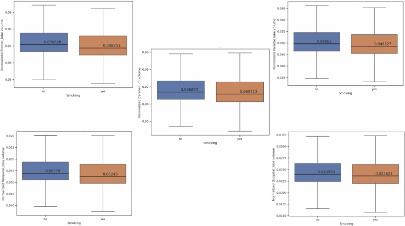
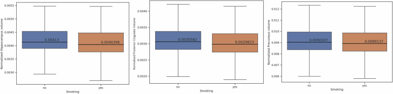
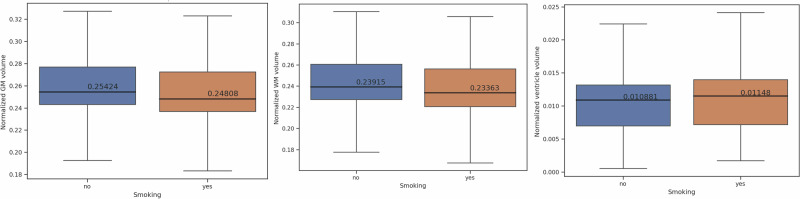
Cigarette smoking is a risk factor for Alzheimer's and vascular dementia, but its impact on brain volume loss, a neurodegeneration biomarker on MRI, is unclear. In total, 10,134 participants from 4 sites were scanned with a whole-body 1.5 T MRI protocol with separate dedicated structural neuroimaging with 3D T1 MPRAGE sequences. Smokers versus non-smokers were compared by gray and white matter volumes normalized to total intracranial volume using a two-tailed t-test. Smokers had lower normalized gray (t = -7.806e+00, p = 6.508e-15) and white matter volumes (t = -7.374e + 00, p = 1.791e-13) compared to non-smokers. Adjusting for age, sex, study site, BMI, and multiple comparisons, higher pack years of smoking predicted volume loss in such regions as total gray matter volume, total white matter volume, temporal lobe, parietal lobe, hippocampus, precuneus, and posterior cingulate. The inclusion and exclusion of BMI from the model suggested an influence of this variable.

 求助内容:
求助内容: 应助结果提醒方式:


