Felicitas J Bijari, Paul Kyu Han, Thibault Marin, Wonil Lee, Yanis Chemli, Inna Gertsenshteyn, Ismaël B G Mounime, Yanis Djebra, Didi Chi, Marc D Normandin, Chao Ma, Georges El Fakhri
{"title":"In vivo 3D myocardial membrane potential mapping in humans using PET/MRI.","authors":"Felicitas J Bijari, Paul Kyu Han, Thibault Marin, Wonil Lee, Yanis Chemli, Inna Gertsenshteyn, Ismaël B G Mounime, Yanis Djebra, Didi Chi, Marc D Normandin, Chao Ma, Georges El Fakhri","doi":"10.1186/s13550-025-01287-7","DOIUrl":null,"url":null,"abstract":"<p><strong>Background: </strong>The mitochondrial membrane potential is a key biophysical parameter of mitochondrial function, which can be useful for the diagnosis and treatment monitoring of various cardiac diseases. We present a non-invasive PET/MR imaging method for 3D myocardial membrane potential mapping in humans.</p><p><strong>Results: </strong>An in vivo PET/MR imaging study was performed in three healthy subjects (1 male and 2 females; 48 ± 29 years old) under a study protocol approved by the local Institutional Review Board (IRB). Written informed consent was obtained from all subjects before participation in the study. The [<sup>18</sup>F](4-Fluorophenyl)triphenylphosphonium ([<sup>18</sup>F]-FTPP<sup>+</sup>) PET tracer was administered using a bolus-plus-infusion protocol (bolus activity of 301.2 ± 7.6 MBq, infusion activity of 90.0 ± 4.9 MBq), where an infusion of 120 min was started shortly after the bolus injection (time of infusion, TOI). Dynamic cardiac PET/MR imaging was performed approximately 20 min after the TOI and continued for 100 min. The extracellular volume fraction mapping was performed via cardiac MR with a free-breathing, 3D cardiac T<sub>1</sub> mapping sequence before and after the contrast agent injection (gadoterate meglumine, 0.1 mmol/kg). A linear tangent space alignment (LTSA) model-based method was used to reconstruct high-frame-rate dynamic images from sparsely sampled (k,t)-space data for T<sub>1</sub>. PET motion correction was performed using two steps of rigid image registration in a multi-resolution fashion, followed by a non-rigid image registration with B-spline transform. The tissue membrane potential was calculated using a kinetic model based on the Nernst equation with myocardial tracer concentration, tracer volume of distribution, and extracellular volume fraction measurements. Fully 3D membrane potential maps were successfully estimated from all three subjects. The estimated whole-heart membrane potentials were - 144.7 ± 3.5 mV, - 160.7 ± 5.3 mV, and - 165.8 ± 3.1 mV for each subject.</p><p><strong>Conclusion: </strong>The proposed method allows 3D myocardial membrane potential mapping in humans in vivo.</p>","PeriodicalId":11611,"journal":{"name":"EJNMMI Research","volume":"15 1","pages":"93"},"PeriodicalIF":3.1000,"publicationDate":"2025-07-26","publicationTypes":"Journal Article","fieldsOfStudy":null,"isOpenAccess":false,"openAccessPdf":"https://www.ncbi.nlm.nih.gov/pmc/articles/PMC12297085/pdf/","citationCount":"0","resultStr":null,"platform":"Semanticscholar","paperid":null,"PeriodicalName":"EJNMMI Research","FirstCategoryId":"3","ListUrlMain":"https://doi.org/10.1186/s13550-025-01287-7","RegionNum":3,"RegionCategory":"医学","ArticlePicture":[],"TitleCN":null,"AbstractTextCN":null,"PMCID":null,"EPubDate":"","PubModel":"","JCR":"Q1","JCRName":"RADIOLOGY, NUCLEAR MEDICINE & MEDICAL IMAGING","Score":null,"Total":0}
引用次数: 0
Abstract
Background: The mitochondrial membrane potential is a key biophysical parameter of mitochondrial function, which can be useful for the diagnosis and treatment monitoring of various cardiac diseases. We present a non-invasive PET/MR imaging method for 3D myocardial membrane potential mapping in humans.
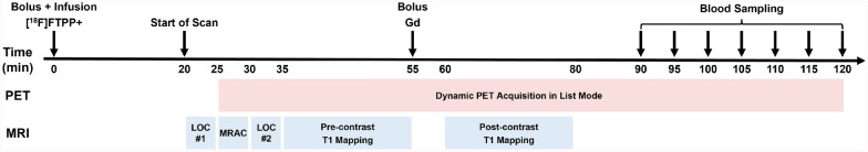
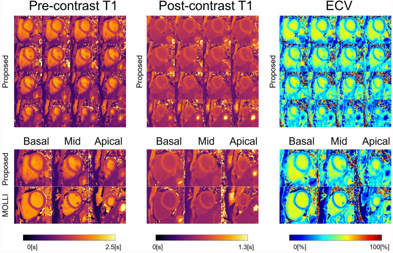
Results: An in vivo PET/MR imaging study was performed in three healthy subjects (1 male and 2 females; 48 ± 29 years old) under a study protocol approved by the local Institutional Review Board (IRB). Written informed consent was obtained from all subjects before participation in the study. The [18F](4-Fluorophenyl)triphenylphosphonium ([18F]-FTPP+) PET tracer was administered using a bolus-plus-infusion protocol (bolus activity of 301.2 ± 7.6 MBq, infusion activity of 90.0 ± 4.9 MBq), where an infusion of 120 min was started shortly after the bolus injection (time of infusion, TOI). Dynamic cardiac PET/MR imaging was performed approximately 20 min after the TOI and continued for 100 min. The extracellular volume fraction mapping was performed via cardiac MR with a free-breathing, 3D cardiac T1 mapping sequence before and after the contrast agent injection (gadoterate meglumine, 0.1 mmol/kg). A linear tangent space alignment (LTSA) model-based method was used to reconstruct high-frame-rate dynamic images from sparsely sampled (k,t)-space data for T1. PET motion correction was performed using two steps of rigid image registration in a multi-resolution fashion, followed by a non-rigid image registration with B-spline transform. The tissue membrane potential was calculated using a kinetic model based on the Nernst equation with myocardial tracer concentration, tracer volume of distribution, and extracellular volume fraction measurements. Fully 3D membrane potential maps were successfully estimated from all three subjects. The estimated whole-heart membrane potentials were - 144.7 ± 3.5 mV, - 160.7 ± 5.3 mV, and - 165.8 ± 3.1 mV for each subject.
Conclusion: The proposed method allows 3D myocardial membrane potential mapping in humans in vivo.
EJNMMI ResearchRADIOLOGY, NUCLEAR MEDICINE & MEDICAL IMAGING&nb-
CiteScore
5.90
自引率
3.10%
发文量
72
审稿时长
13 weeks
期刊介绍:
EJNMMI Research publishes new basic, translational and clinical research in the field of nuclear medicine and molecular imaging. Regular features include original research articles, rapid communication of preliminary data on innovative research, interesting case reports, editorials, and letters to the editor. Educational articles on basic sciences, fundamental aspects and controversy related to pre-clinical and clinical research or ethical aspects of research are also welcome. Timely reviews provide updates on current applications, issues in imaging research and translational aspects of nuclear medicine and molecular imaging technologies.
The main emphasis is placed on the development of targeted imaging with radiopharmaceuticals within the broader context of molecular probes to enhance understanding and characterisation of the complex biological processes underlying disease and to develop, test and guide new treatment modalities, including radionuclide therapy.


 求助内容:
求助内容: 应助结果提醒方式:


