Hande Nur Taşdemir Batir, Hatice Güler, Burcu Kamaşak Arpaçay, İzzet Ökçesiz, Halil Dönmez, Güven Kahriman
{"title":"Morphometric Analysis of Subaxial Cervical Vertebra Pedicles in the Turkish Population.","authors":"Hande Nur Taşdemir Batir, Hatice Güler, Burcu Kamaşak Arpaçay, İzzet Ökçesiz, Halil Dönmez, Güven Kahriman","doi":"10.3390/tomography11070079","DOIUrl":null,"url":null,"abstract":"<p><strong>Background/objectives: </strong>One of the surgical interventions applied in the cervical region is the pedicle screw method. The cervical pedicle screw is stronger than any other screw method; however, use of the cervical pedicle screw is limited due to the variability in the anatomy of the cervical vertebrae and the risks to the neurological and vascular structures in this region. This study aimed to determine the morphological features of subaxial cervical vertebrae of the adult Turkish population and to provide guidance for the pedicle screwing method.</p><p><strong>Methods: </strong>In our study, pedicle analyses were examined in the subaxial neck vertebrae of a total of 60 patients, 30 male and 30 female, using computed tomography images. In subaxial vertebrae (C3-C7), bilateral pedicle width, pedicle axis length, pedicle transverse angle, sagittal and transverse diameter of vertebral foramen, and the distance between two pedicles were measured.</p><p><strong>Results: </strong>Pedicle widths that did not fit the commonly used 3.5 mm pedicle screw were detected in both male and female patients. The mean bilateral pedicle width in male patients was found to be greater than in female patients. When the parameter results were compared according to the levels, it was found that the pedicle width, pedicle axis length, transverse diameter, and the distance between the two pedicles increased statistically significantly.</p><p><strong>Conclusions: </strong>We think that the data obtained from the study will help determine the appropriate screwing (screw selection) in subaxial vertebra pedicle surgery and increase the success of the surgical procedure.</p>","PeriodicalId":51330,"journal":{"name":"Tomography","volume":"11 7","pages":""},"PeriodicalIF":2.2000,"publicationDate":"2025-07-04","publicationTypes":"Journal Article","fieldsOfStudy":null,"isOpenAccess":false,"openAccessPdf":"https://www.ncbi.nlm.nih.gov/pmc/articles/PMC12298989/pdf/","citationCount":"0","resultStr":null,"platform":"Semanticscholar","paperid":null,"PeriodicalName":"Tomography","FirstCategoryId":"3","ListUrlMain":"https://doi.org/10.3390/tomography11070079","RegionNum":4,"RegionCategory":"医学","ArticlePicture":[],"TitleCN":null,"AbstractTextCN":null,"PMCID":null,"EPubDate":"","PubModel":"","JCR":"Q2","JCRName":"RADIOLOGY, NUCLEAR MEDICINE & MEDICAL IMAGING","Score":null,"Total":0}
引用次数: 0
Abstract
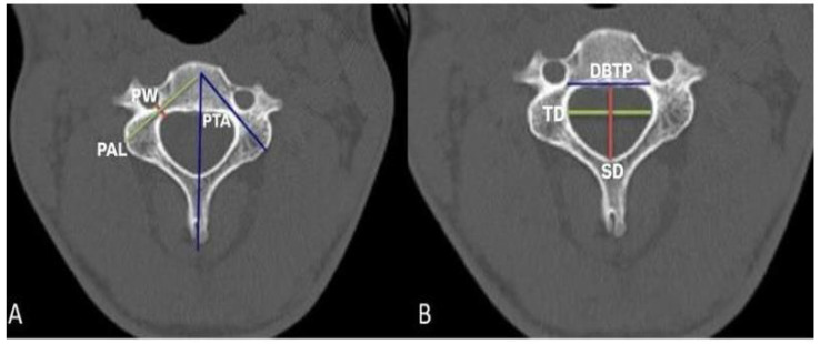
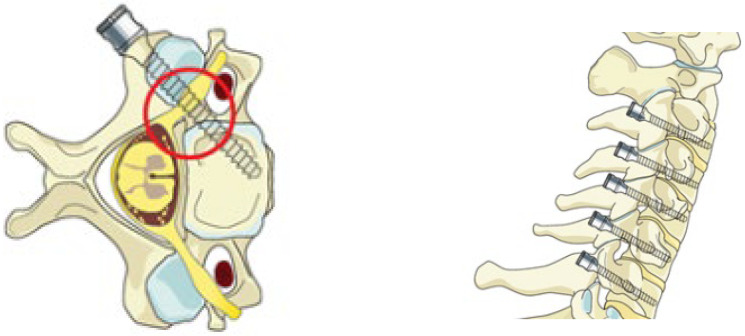
Background/objectives: One of the surgical interventions applied in the cervical region is the pedicle screw method. The cervical pedicle screw is stronger than any other screw method; however, use of the cervical pedicle screw is limited due to the variability in the anatomy of the cervical vertebrae and the risks to the neurological and vascular structures in this region. This study aimed to determine the morphological features of subaxial cervical vertebrae of the adult Turkish population and to provide guidance for the pedicle screwing method.
Methods: In our study, pedicle analyses were examined in the subaxial neck vertebrae of a total of 60 patients, 30 male and 30 female, using computed tomography images. In subaxial vertebrae (C3-C7), bilateral pedicle width, pedicle axis length, pedicle transverse angle, sagittal and transverse diameter of vertebral foramen, and the distance between two pedicles were measured.
Results: Pedicle widths that did not fit the commonly used 3.5 mm pedicle screw were detected in both male and female patients. The mean bilateral pedicle width in male patients was found to be greater than in female patients. When the parameter results were compared according to the levels, it was found that the pedicle width, pedicle axis length, transverse diameter, and the distance between the two pedicles increased statistically significantly.
Conclusions: We think that the data obtained from the study will help determine the appropriate screwing (screw selection) in subaxial vertebra pedicle surgery and increase the success of the surgical procedure.
TomographyMedicine-Radiology, Nuclear Medicine and Imaging
CiteScore
2.70
自引率
10.50%
发文量
222
期刊介绍:
TomographyTM publishes basic (technical and pre-clinical) and clinical scientific articles which involve the advancement of imaging technologies. Tomography encompasses studies that use single or multiple imaging modalities including for example CT, US, PET, SPECT, MR and hyperpolarization technologies, as well as optical modalities (i.e. bioluminescence, photoacoustic, endomicroscopy, fiber optic imaging and optical computed tomography) in basic sciences, engineering, preclinical and clinical medicine.
Tomography also welcomes studies involving exploration and refinement of contrast mechanisms and image-derived metrics within and across modalities toward the development of novel imaging probes for image-based feedback and intervention. The use of imaging in biology and medicine provides unparalleled opportunities to noninvasively interrogate tissues to obtain real-time dynamic and quantitative information required for diagnosis and response to interventions and to follow evolving pathological conditions. As multi-modal studies and the complexities of imaging technologies themselves are ever increasing to provide advanced information to scientists and clinicians.
Tomography provides a unique publication venue allowing investigators the opportunity to more precisely communicate integrated findings related to the diverse and heterogeneous features associated with underlying anatomical, physiological, functional, metabolic and molecular genetic activities of normal and diseased tissue. Thus Tomography publishes peer-reviewed articles which involve the broad use of imaging of any tissue and disease type including both preclinical and clinical investigations. In addition, hardware/software along with chemical and molecular probe advances are welcome as they are deemed to significantly contribute towards the long-term goal of improving the overall impact of imaging on scientific and clinical discovery.

 求助内容:
求助内容: 应助结果提醒方式:


