Giuseppe Sarti, Giovanni Barbato, Francesco Tiralongo, Gianpaolo Santini, Francesco Arienzo, Davide Nilo, Fabio Tortora, Alfonso Reginelli, Rosita Comune, Maria Borrelli, Stefania Tamburrini, Antonio Basile, Mariano Scaglione
{"title":"Endovascular Treatment of Extracranial Arteriovenous Malformations: A Retrospective Monocentric Case-Series Study.","authors":"Giuseppe Sarti, Giovanni Barbato, Francesco Tiralongo, Gianpaolo Santini, Francesco Arienzo, Davide Nilo, Fabio Tortora, Alfonso Reginelli, Rosita Comune, Maria Borrelli, Stefania Tamburrini, Antonio Basile, Mariano Scaglione","doi":"10.3390/tomography11070075","DOIUrl":null,"url":null,"abstract":"<p><strong>Background: </strong>Extracranial arteriovenous malformations (AVMs) are rare congenital vascular anomalies that often require endovascular treatment due to symptoms such as pain, bleeding, or functional impairment. Endovascular strategies include arterial, venous, or combined embolization approaches; however, recurrence remains a major challenge. We retrospectively evaluate the technical success, safety, and clinical outcomes of arterial-only versus combined arterial and venous embolization for the treatment of extracranial AVMs.</p><p><strong>Materials and methods: </strong>This single-center retrospective study included 14 patients (mean age 31.8 ± 21.7 years; 64% female) with symptomatic extracranial AVMs (Schobinger stage II) treated between 2017 and 2023. AVMs were classified angiographically (Yakes classification) and treated with embolization via arterial or combined access routes. The primary endpoint was technical success (defined as angiographic nidus occlusion), while secondary endpoints included clinical recurrence and procedure-related complications. Follow-up included clinical and Doppler ultrasound assessments.</p><p><strong>Results: </strong>Nine patients (64%) underwent arterial embolization alone; five (36%) received combined arterial and venous embolization, including Lauromacrogol injection via direct puncture. Technical success was achieved in all cases (100%). Clinical recurrence occurred in two patients (14%), both from the arterial-only group. One major complication (tongue ischemia) occurred in a single patient (7%). No complications or recurrences were observed in the combined treatment group. Statistical analysis showed no significant difference in recurrence or complication rates between groups.</p>","PeriodicalId":51330,"journal":{"name":"Tomography","volume":"11 7","pages":""},"PeriodicalIF":2.2000,"publicationDate":"2025-06-26","publicationTypes":"Journal Article","fieldsOfStudy":null,"isOpenAccess":false,"openAccessPdf":"https://www.ncbi.nlm.nih.gov/pmc/articles/PMC12300876/pdf/","citationCount":"0","resultStr":null,"platform":"Semanticscholar","paperid":null,"PeriodicalName":"Tomography","FirstCategoryId":"3","ListUrlMain":"https://doi.org/10.3390/tomography11070075","RegionNum":4,"RegionCategory":"医学","ArticlePicture":[],"TitleCN":null,"AbstractTextCN":null,"PMCID":null,"EPubDate":"","PubModel":"","JCR":"Q2","JCRName":"RADIOLOGY, NUCLEAR MEDICINE & MEDICAL IMAGING","Score":null,"Total":0}
引用次数: 0
Abstract
Background: Extracranial arteriovenous malformations (AVMs) are rare congenital vascular anomalies that often require endovascular treatment due to symptoms such as pain, bleeding, or functional impairment. Endovascular strategies include arterial, venous, or combined embolization approaches; however, recurrence remains a major challenge. We retrospectively evaluate the technical success, safety, and clinical outcomes of arterial-only versus combined arterial and venous embolization for the treatment of extracranial AVMs.
Materials and methods: This single-center retrospective study included 14 patients (mean age 31.8 ± 21.7 years; 64% female) with symptomatic extracranial AVMs (Schobinger stage II) treated between 2017 and 2023. AVMs were classified angiographically (Yakes classification) and treated with embolization via arterial or combined access routes. The primary endpoint was technical success (defined as angiographic nidus occlusion), while secondary endpoints included clinical recurrence and procedure-related complications. Follow-up included clinical and Doppler ultrasound assessments.
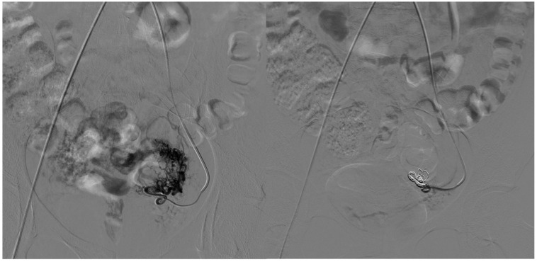
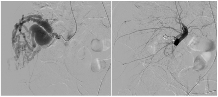
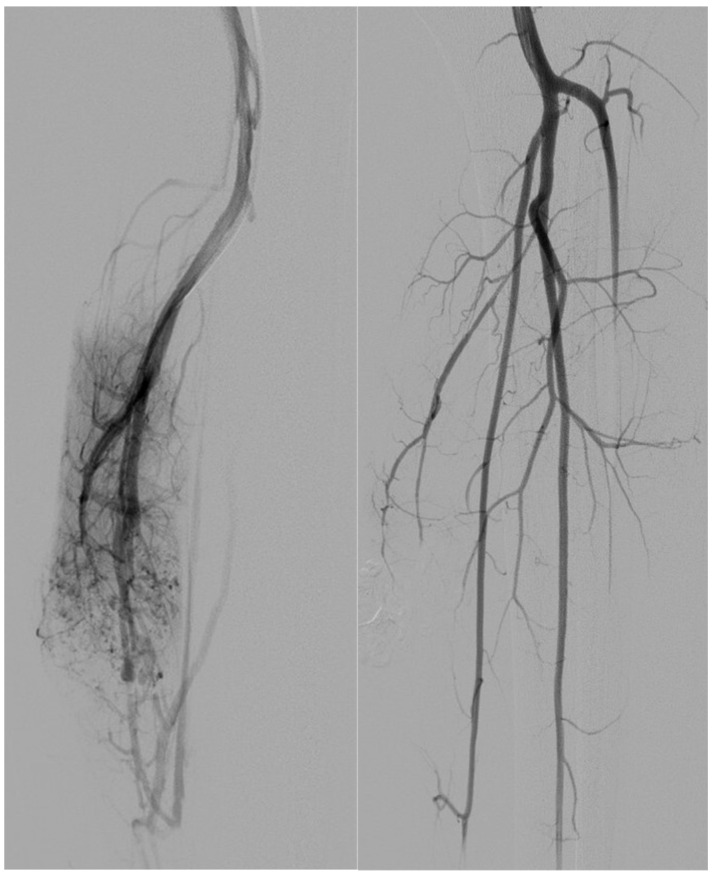
Results: Nine patients (64%) underwent arterial embolization alone; five (36%) received combined arterial and venous embolization, including Lauromacrogol injection via direct puncture. Technical success was achieved in all cases (100%). Clinical recurrence occurred in two patients (14%), both from the arterial-only group. One major complication (tongue ischemia) occurred in a single patient (7%). No complications or recurrences were observed in the combined treatment group. Statistical analysis showed no significant difference in recurrence or complication rates between groups.
TomographyMedicine-Radiology, Nuclear Medicine and Imaging
CiteScore
2.70
自引率
10.50%
发文量
222
期刊介绍:
TomographyTM publishes basic (technical and pre-clinical) and clinical scientific articles which involve the advancement of imaging technologies. Tomography encompasses studies that use single or multiple imaging modalities including for example CT, US, PET, SPECT, MR and hyperpolarization technologies, as well as optical modalities (i.e. bioluminescence, photoacoustic, endomicroscopy, fiber optic imaging and optical computed tomography) in basic sciences, engineering, preclinical and clinical medicine.
Tomography also welcomes studies involving exploration and refinement of contrast mechanisms and image-derived metrics within and across modalities toward the development of novel imaging probes for image-based feedback and intervention. The use of imaging in biology and medicine provides unparalleled opportunities to noninvasively interrogate tissues to obtain real-time dynamic and quantitative information required for diagnosis and response to interventions and to follow evolving pathological conditions. As multi-modal studies and the complexities of imaging technologies themselves are ever increasing to provide advanced information to scientists and clinicians.
Tomography provides a unique publication venue allowing investigators the opportunity to more precisely communicate integrated findings related to the diverse and heterogeneous features associated with underlying anatomical, physiological, functional, metabolic and molecular genetic activities of normal and diseased tissue. Thus Tomography publishes peer-reviewed articles which involve the broad use of imaging of any tissue and disease type including both preclinical and clinical investigations. In addition, hardware/software along with chemical and molecular probe advances are welcome as they are deemed to significantly contribute towards the long-term goal of improving the overall impact of imaging on scientific and clinical discovery.

 求助内容:
求助内容: 应助结果提醒方式:


