Randy Guzmán Gómez, Guadalupe Lopez Lopez, Victor M Alvarado, Froylan Lopez Lopez, Eréndira Esqueda Cisneros, Hazel López Moreno
{"title":"Deep Learning Approaches for Automated Prediction of Treatment Response in Non-Small-Cell Lung Cancer Patients Based on CT and PET Imaging.","authors":"Randy Guzmán Gómez, Guadalupe Lopez Lopez, Victor M Alvarado, Froylan Lopez Lopez, Eréndira Esqueda Cisneros, Hazel López Moreno","doi":"10.3390/tomography11070078","DOIUrl":null,"url":null,"abstract":"<p><p>The rapid growth of artificial intelligence, particularly in the field of deep learning, has opened up new advances in analyzing and processing large and complex datasets. Prospects and emerging trends in this area engage the development of methods, techniques, and algorithms to build autonomous systems that perform tasks with minimal human action. In medical practice, radiological imaging technologies systematically boost progress in the clinical monitoring of cancer through the information that can be analyzed in these images. This review gives insight into deep learning-based approaches that strengthen the assessment of the response to the treatment of non-small-cell lung cancer. This systematic survey delves into the various approaches to morphological and metabolic changes observed in computerized tomography (CT) and positron emission tomography (PET) imaging. We highlight the challenges and opportunities for feasible integration of deep learning computer-based tools in evaluating treatments in lung cancer patients, after which CT and PET-based strategies are contrasted. The investigated deep learning methods are organized and described as instruments for classification, clustering, and prediction, which can contribute to the design of automated and objective assessment of lung tumor responses to treatments.</p>","PeriodicalId":51330,"journal":{"name":"Tomography","volume":"11 7","pages":""},"PeriodicalIF":2.2000,"publicationDate":"2025-06-30","publicationTypes":"Journal Article","fieldsOfStudy":null,"isOpenAccess":false,"openAccessPdf":"https://www.ncbi.nlm.nih.gov/pmc/articles/PMC12298732/pdf/","citationCount":"0","resultStr":null,"platform":"Semanticscholar","paperid":null,"PeriodicalName":"Tomography","FirstCategoryId":"3","ListUrlMain":"https://doi.org/10.3390/tomography11070078","RegionNum":4,"RegionCategory":"医学","ArticlePicture":[],"TitleCN":null,"AbstractTextCN":null,"PMCID":null,"EPubDate":"","PubModel":"","JCR":"Q2","JCRName":"RADIOLOGY, NUCLEAR MEDICINE & MEDICAL IMAGING","Score":null,"Total":0}
引用次数: 0
Abstract
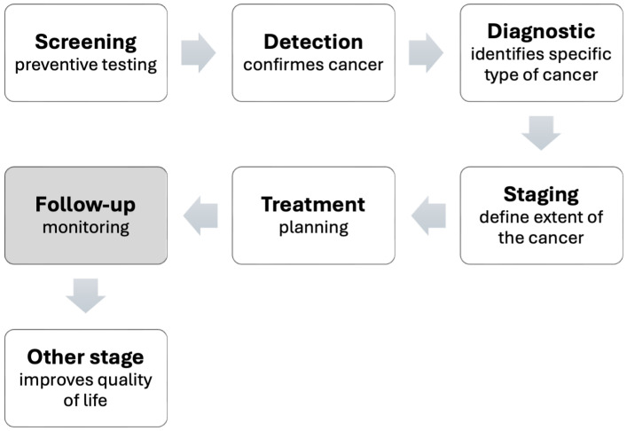
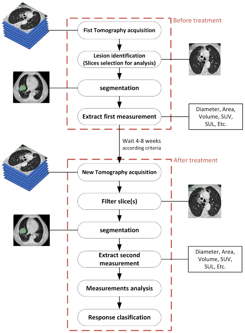
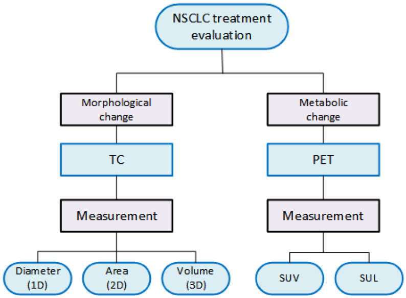
The rapid growth of artificial intelligence, particularly in the field of deep learning, has opened up new advances in analyzing and processing large and complex datasets. Prospects and emerging trends in this area engage the development of methods, techniques, and algorithms to build autonomous systems that perform tasks with minimal human action. In medical practice, radiological imaging technologies systematically boost progress in the clinical monitoring of cancer through the information that can be analyzed in these images. This review gives insight into deep learning-based approaches that strengthen the assessment of the response to the treatment of non-small-cell lung cancer. This systematic survey delves into the various approaches to morphological and metabolic changes observed in computerized tomography (CT) and positron emission tomography (PET) imaging. We highlight the challenges and opportunities for feasible integration of deep learning computer-based tools in evaluating treatments in lung cancer patients, after which CT and PET-based strategies are contrasted. The investigated deep learning methods are organized and described as instruments for classification, clustering, and prediction, which can contribute to the design of automated and objective assessment of lung tumor responses to treatments.
TomographyMedicine-Radiology, Nuclear Medicine and Imaging
CiteScore
2.70
自引率
10.50%
发文量
222
期刊介绍:
TomographyTM publishes basic (technical and pre-clinical) and clinical scientific articles which involve the advancement of imaging technologies. Tomography encompasses studies that use single or multiple imaging modalities including for example CT, US, PET, SPECT, MR and hyperpolarization technologies, as well as optical modalities (i.e. bioluminescence, photoacoustic, endomicroscopy, fiber optic imaging and optical computed tomography) in basic sciences, engineering, preclinical and clinical medicine.
Tomography also welcomes studies involving exploration and refinement of contrast mechanisms and image-derived metrics within and across modalities toward the development of novel imaging probes for image-based feedback and intervention. The use of imaging in biology and medicine provides unparalleled opportunities to noninvasively interrogate tissues to obtain real-time dynamic and quantitative information required for diagnosis and response to interventions and to follow evolving pathological conditions. As multi-modal studies and the complexities of imaging technologies themselves are ever increasing to provide advanced information to scientists and clinicians.
Tomography provides a unique publication venue allowing investigators the opportunity to more precisely communicate integrated findings related to the diverse and heterogeneous features associated with underlying anatomical, physiological, functional, metabolic and molecular genetic activities of normal and diseased tissue. Thus Tomography publishes peer-reviewed articles which involve the broad use of imaging of any tissue and disease type including both preclinical and clinical investigations. In addition, hardware/software along with chemical and molecular probe advances are welcome as they are deemed to significantly contribute towards the long-term goal of improving the overall impact of imaging on scientific and clinical discovery.

 求助内容:
求助内容: 应助结果提醒方式:


