Victória Geisa Brito de Oliveira, Polyane Mazucatto Queiroz, Alessandra Rocha Simões, Mônica Ghislaine Oliveira Alves, Maria Aparecida Neves Jardini, André Luiz Ferreira Costa, Sérgio Lucio Pereira de Castro Lopes
{"title":"Voxel Size and Field of View Influence on Periodontal Bone Assessment Using Four CBCT Systems: An Experimental Ex Vivo Analysis.","authors":"Victória Geisa Brito de Oliveira, Polyane Mazucatto Queiroz, Alessandra Rocha Simões, Mônica Ghislaine Oliveira Alves, Maria Aparecida Neves Jardini, André Luiz Ferreira Costa, Sérgio Lucio Pereira de Castro Lopes","doi":"10.3390/tomography11070074","DOIUrl":null,"url":null,"abstract":"<p><strong>Objective: </strong>This ex vivo study aimed to evaluate the influence of different acquisition protocols, combining voxel size and field of view (FOV), across four cone-beam computed tomography (CBCT) systems, on the accuracy of alveolar bone level measurements for periodontal assessment.</p><p><strong>Materials and methods: </strong>A dry human mandible was used, with standardized radiopaque markers placed on the cementoenamel junction (CEJ) of the buccal-mesial and buccal-distal aspects of teeth 34 and 43. CBCT scans were performed using four systems-Veraview<sup>®</sup> X800, OP300 Pro<sup>®</sup>, I-CAT Next Generation<sup>®</sup>, and Orthophos XG<sup>®</sup>-applying various combinations of field of view (FOV) and voxel resolution available in each device. Reference measurements were obtained in situ using a digital caliper. CBCT images were exported in DICOM format and analyzed with OnDemand3D software (version 4.6) to obtain paracoronal sections. Linear measurements from the CEJ to the alveolar crest were recorded in triplicate and compared to the gold standard using ANOVA and the Dunnett test (α = 0.05).</p><p><strong>Results: </strong>Protocols with smaller voxel sizes and limited FOVs generally yielded measurements closer to the gold standard. However, some larger-FOV protocols with intermediate voxel sizes also achieved comparable accuracy. Among the systems, the I-CAT showed lower agreement within in situ measurements, while others demonstrated reliable performance depending on the acquisition parameters.</p><p><strong>Conclusions: </strong>The findings suggest that CBCT protocols with smaller voxel sizes and reduced FOVs can enhance measurement accuracy in periodontal bone assessments. Nevertheless, intermediate protocols may offer a balance between diagnostic quality and radiation exposure, aligning with the ALADA principle. This study reinforces the need for standardized acquisition parameters tailored to periodontal imaging.</p>","PeriodicalId":51330,"journal":{"name":"Tomography","volume":"11 7","pages":""},"PeriodicalIF":2.2000,"publicationDate":"2025-06-25","publicationTypes":"Journal Article","fieldsOfStudy":null,"isOpenAccess":false,"openAccessPdf":"https://www.ncbi.nlm.nih.gov/pmc/articles/PMC12300106/pdf/","citationCount":"0","resultStr":null,"platform":"Semanticscholar","paperid":null,"PeriodicalName":"Tomography","FirstCategoryId":"3","ListUrlMain":"https://doi.org/10.3390/tomography11070074","RegionNum":4,"RegionCategory":"医学","ArticlePicture":[],"TitleCN":null,"AbstractTextCN":null,"PMCID":null,"EPubDate":"","PubModel":"","JCR":"Q2","JCRName":"RADIOLOGY, NUCLEAR MEDICINE & MEDICAL IMAGING","Score":null,"Total":0}
引用次数: 0
Abstract
Objective: This ex vivo study aimed to evaluate the influence of different acquisition protocols, combining voxel size and field of view (FOV), across four cone-beam computed tomography (CBCT) systems, on the accuracy of alveolar bone level measurements for periodontal assessment.
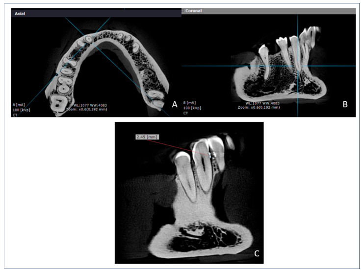
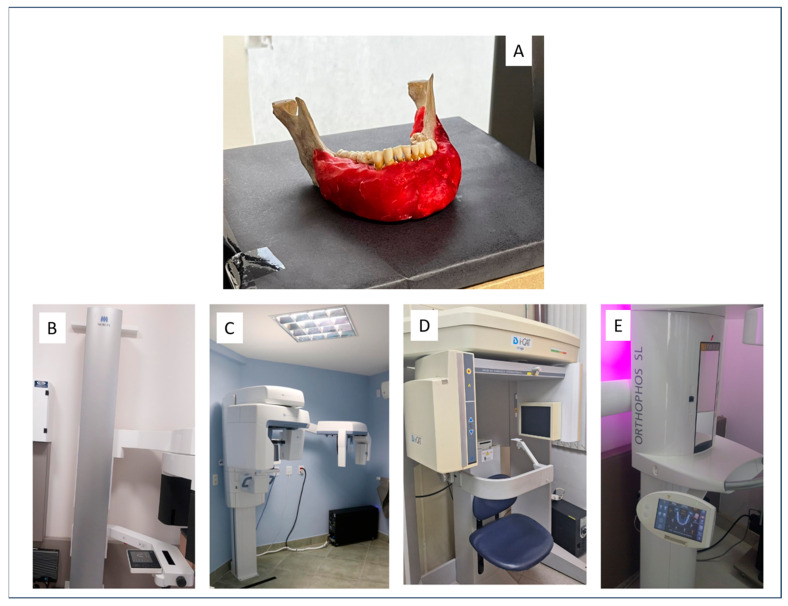
Materials and methods: A dry human mandible was used, with standardized radiopaque markers placed on the cementoenamel junction (CEJ) of the buccal-mesial and buccal-distal aspects of teeth 34 and 43. CBCT scans were performed using four systems-Veraview® X800, OP300 Pro®, I-CAT Next Generation®, and Orthophos XG®-applying various combinations of field of view (FOV) and voxel resolution available in each device. Reference measurements were obtained in situ using a digital caliper. CBCT images were exported in DICOM format and analyzed with OnDemand3D software (version 4.6) to obtain paracoronal sections. Linear measurements from the CEJ to the alveolar crest were recorded in triplicate and compared to the gold standard using ANOVA and the Dunnett test (α = 0.05).
Results: Protocols with smaller voxel sizes and limited FOVs generally yielded measurements closer to the gold standard. However, some larger-FOV protocols with intermediate voxel sizes also achieved comparable accuracy. Among the systems, the I-CAT showed lower agreement within in situ measurements, while others demonstrated reliable performance depending on the acquisition parameters.
Conclusions: The findings suggest that CBCT protocols with smaller voxel sizes and reduced FOVs can enhance measurement accuracy in periodontal bone assessments. Nevertheless, intermediate protocols may offer a balance between diagnostic quality and radiation exposure, aligning with the ALADA principle. This study reinforces the need for standardized acquisition parameters tailored to periodontal imaging.
TomographyMedicine-Radiology, Nuclear Medicine and Imaging
CiteScore
2.70
自引率
10.50%
发文量
222
期刊介绍:
TomographyTM publishes basic (technical and pre-clinical) and clinical scientific articles which involve the advancement of imaging technologies. Tomography encompasses studies that use single or multiple imaging modalities including for example CT, US, PET, SPECT, MR and hyperpolarization technologies, as well as optical modalities (i.e. bioluminescence, photoacoustic, endomicroscopy, fiber optic imaging and optical computed tomography) in basic sciences, engineering, preclinical and clinical medicine.
Tomography also welcomes studies involving exploration and refinement of contrast mechanisms and image-derived metrics within and across modalities toward the development of novel imaging probes for image-based feedback and intervention. The use of imaging in biology and medicine provides unparalleled opportunities to noninvasively interrogate tissues to obtain real-time dynamic and quantitative information required for diagnosis and response to interventions and to follow evolving pathological conditions. As multi-modal studies and the complexities of imaging technologies themselves are ever increasing to provide advanced information to scientists and clinicians.
Tomography provides a unique publication venue allowing investigators the opportunity to more precisely communicate integrated findings related to the diverse and heterogeneous features associated with underlying anatomical, physiological, functional, metabolic and molecular genetic activities of normal and diseased tissue. Thus Tomography publishes peer-reviewed articles which involve the broad use of imaging of any tissue and disease type including both preclinical and clinical investigations. In addition, hardware/software along with chemical and molecular probe advances are welcome as they are deemed to significantly contribute towards the long-term goal of improving the overall impact of imaging on scientific and clinical discovery.

 求助内容:
求助内容: 应助结果提醒方式:


