{"title":"Qualitative and Quantitative Computed Tomography Analyses of Lung Adenocarcinoma for Predicting Spread Through Air Spaces.","authors":"Fumi Kameda, Yoshie Kunihiro, Masahiro Tanabe, Masatoshi Nakashima, Taiga Kobayashi, Toshiki Tanaka, Yoshinobu Hoshii, Katsuyoshi Ito","doi":"10.3390/tomography11070076","DOIUrl":null,"url":null,"abstract":"<p><strong>Background/objectives: </strong>Spread through air spaces (STAS) is defined as the spread of tumor cells into the parenchymal alveolar space beyond the margins of the main tumor, and it is associated with worse clinical outcomes in resected lung adenocarcinoma. This study aimed to evaluate the preoperative computed tomography (CT) findings of primary lung adenocarcinoma in surgically resected T1 cases and to compare CT findings with and without STAS.</p><p><strong>Methods: </strong>A total of 145 patients were included in this study. The following factors were evaluated on CT images: nodule type (pure ground-glass nodule [GGN], part-solid nodule, or solid nodule), margin (smooth or irregular), the presence of lobulation, spicula, cavity, calcification, central low attenuation, peripheral opacity (well-defined or ill-defined), air bronchogram, satellite lesions, pleural retraction, pulmonary emphysema, and interstitial pneumonia; CT values (maximum, minimum, and mean); volume (tumor and solid component); and diameter (tumor and solid component). CT criteria were compared between the presence and absence of STAS.</p><p><strong>Results: </strong>Lobulation and central low attenuation were significantly more frequent in patients with STAS (<i>p</i> < 0.05). The mean CT value, and the volume, rate, and diameter of the solid component were significantly larger in cases with STAS (<i>p</i> < 0.05). A multiple logistic regression analysis identified central low attenuation as an indicator of the presence of STAS (<i>p</i> < 0.001; odds ratio, 3.993; 95% confidence interval, 1.993-8.001).</p><p><strong>Conclusions: </strong>Quantitative and qualitative analyses are useful for differentiating between the presence and absence of STAS.</p>","PeriodicalId":51330,"journal":{"name":"Tomography","volume":"11 7","pages":""},"PeriodicalIF":2.2000,"publicationDate":"2025-06-27","publicationTypes":"Journal Article","fieldsOfStudy":null,"isOpenAccess":false,"openAccessPdf":"https://www.ncbi.nlm.nih.gov/pmc/articles/PMC12298125/pdf/","citationCount":"0","resultStr":null,"platform":"Semanticscholar","paperid":null,"PeriodicalName":"Tomography","FirstCategoryId":"3","ListUrlMain":"https://doi.org/10.3390/tomography11070076","RegionNum":4,"RegionCategory":"医学","ArticlePicture":[],"TitleCN":null,"AbstractTextCN":null,"PMCID":null,"EPubDate":"","PubModel":"","JCR":"Q2","JCRName":"RADIOLOGY, NUCLEAR MEDICINE & MEDICAL IMAGING","Score":null,"Total":0}
引用次数: 0
Abstract
Background/objectives: Spread through air spaces (STAS) is defined as the spread of tumor cells into the parenchymal alveolar space beyond the margins of the main tumor, and it is associated with worse clinical outcomes in resected lung adenocarcinoma. This study aimed to evaluate the preoperative computed tomography (CT) findings of primary lung adenocarcinoma in surgically resected T1 cases and to compare CT findings with and without STAS.
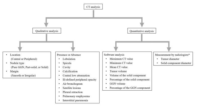
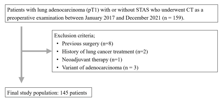
Methods: A total of 145 patients were included in this study. The following factors were evaluated on CT images: nodule type (pure ground-glass nodule [GGN], part-solid nodule, or solid nodule), margin (smooth or irregular), the presence of lobulation, spicula, cavity, calcification, central low attenuation, peripheral opacity (well-defined or ill-defined), air bronchogram, satellite lesions, pleural retraction, pulmonary emphysema, and interstitial pneumonia; CT values (maximum, minimum, and mean); volume (tumor and solid component); and diameter (tumor and solid component). CT criteria were compared between the presence and absence of STAS.
Results: Lobulation and central low attenuation were significantly more frequent in patients with STAS (p < 0.05). The mean CT value, and the volume, rate, and diameter of the solid component were significantly larger in cases with STAS (p < 0.05). A multiple logistic regression analysis identified central low attenuation as an indicator of the presence of STAS (p < 0.001; odds ratio, 3.993; 95% confidence interval, 1.993-8.001).
Conclusions: Quantitative and qualitative analyses are useful for differentiating between the presence and absence of STAS.
TomographyMedicine-Radiology, Nuclear Medicine and Imaging
CiteScore
2.70
自引率
10.50%
发文量
222
期刊介绍:
TomographyTM publishes basic (technical and pre-clinical) and clinical scientific articles which involve the advancement of imaging technologies. Tomography encompasses studies that use single or multiple imaging modalities including for example CT, US, PET, SPECT, MR and hyperpolarization technologies, as well as optical modalities (i.e. bioluminescence, photoacoustic, endomicroscopy, fiber optic imaging and optical computed tomography) in basic sciences, engineering, preclinical and clinical medicine.
Tomography also welcomes studies involving exploration and refinement of contrast mechanisms and image-derived metrics within and across modalities toward the development of novel imaging probes for image-based feedback and intervention. The use of imaging in biology and medicine provides unparalleled opportunities to noninvasively interrogate tissues to obtain real-time dynamic and quantitative information required for diagnosis and response to interventions and to follow evolving pathological conditions. As multi-modal studies and the complexities of imaging technologies themselves are ever increasing to provide advanced information to scientists and clinicians.
Tomography provides a unique publication venue allowing investigators the opportunity to more precisely communicate integrated findings related to the diverse and heterogeneous features associated with underlying anatomical, physiological, functional, metabolic and molecular genetic activities of normal and diseased tissue. Thus Tomography publishes peer-reviewed articles which involve the broad use of imaging of any tissue and disease type including both preclinical and clinical investigations. In addition, hardware/software along with chemical and molecular probe advances are welcome as they are deemed to significantly contribute towards the long-term goal of improving the overall impact of imaging on scientific and clinical discovery.

 求助内容:
求助内容: 应助结果提醒方式:


