Implantation of an Artificial Intelligence Denoising Algorithm Using SubtlePET™ with Various Radiotracers: 18F-FDG, 68Ga PSMA-11 and 18F-FDOPA, Impact on the Technologist Radiation Doses.
IF 2.7 Q3 IMAGING SCIENCE & PHOTOGRAPHIC TECHNOLOGY
Jules Zhang-Yin, Octavian Dragusin, Paul Jonard, Christian Picard, Justine Grangeret, Christopher Bonnier, Philippe P Leveque, Joel Aerts, Olivier Schaeffer
{"title":"Implantation of an Artificial Intelligence Denoising Algorithm Using SubtlePET™ with Various Radiotracers: 18F-FDG, 68Ga PSMA-11 and 18F-FDOPA, Impact on the Technologist Radiation Doses.","authors":"Jules Zhang-Yin, Octavian Dragusin, Paul Jonard, Christian Picard, Justine Grangeret, Christopher Bonnier, Philippe P Leveque, Joel Aerts, Olivier Schaeffer","doi":"10.3390/jimaging11070234","DOIUrl":null,"url":null,"abstract":"<p><p>This study assesses the clinical deployment of SubtlePET™, a commercial AI-based denoising algorithm, across three radiotracers-<sup>18</sup>F-FDG, <sup>68</sup>Ga-PSMA-11, and <sup>18</sup>F-FDOPA-with the goal of improving image quality while reducing injected activity, technologist radiation exposure, and scan time. A retrospective analysis on a digital PET/CT system showed that SubtlePET™ enabled dose reductions exceeding 33% and time savings of over 25%. AI-enhanced images were rated interpretable in 100% of cases versus 65% for standard low-dose reconstructions. Notably, 85% of AI-enhanced scans received the maximum Likert quality score (5/5), indicating excellent diagnostic confidence and noise suppression, compared to only 50% with conventional reconstruction. The quantitative image quality improved significantly across all tracers, with SNR and CNR gains of 50-70%. Radiotracer dose reductions were particularly substantial in low-BMI patients (up to 41% for FDG), and the technologist exposure decreased for high-exposure roles. The daily patient throughput increased by an average of 4.84 cases. These findings support the robust integration of SubtlePET™ into routine clinical PET practice, offering improved efficiency, safety, and image quality without compromising lesion detectability.</p>","PeriodicalId":37035,"journal":{"name":"Journal of Imaging","volume":"11 7","pages":""},"PeriodicalIF":2.7000,"publicationDate":"2025-07-11","publicationTypes":"Journal Article","fieldsOfStudy":null,"isOpenAccess":false,"openAccessPdf":"https://www.ncbi.nlm.nih.gov/pmc/articles/PMC12295822/pdf/","citationCount":"0","resultStr":null,"platform":"Semanticscholar","paperid":null,"PeriodicalName":"Journal of Imaging","FirstCategoryId":"1085","ListUrlMain":"https://doi.org/10.3390/jimaging11070234","RegionNum":0,"RegionCategory":null,"ArticlePicture":[],"TitleCN":null,"AbstractTextCN":null,"PMCID":null,"EPubDate":"","PubModel":"","JCR":"Q3","JCRName":"IMAGING SCIENCE & PHOTOGRAPHIC TECHNOLOGY","Score":null,"Total":0}
引用次数: 0
Abstract
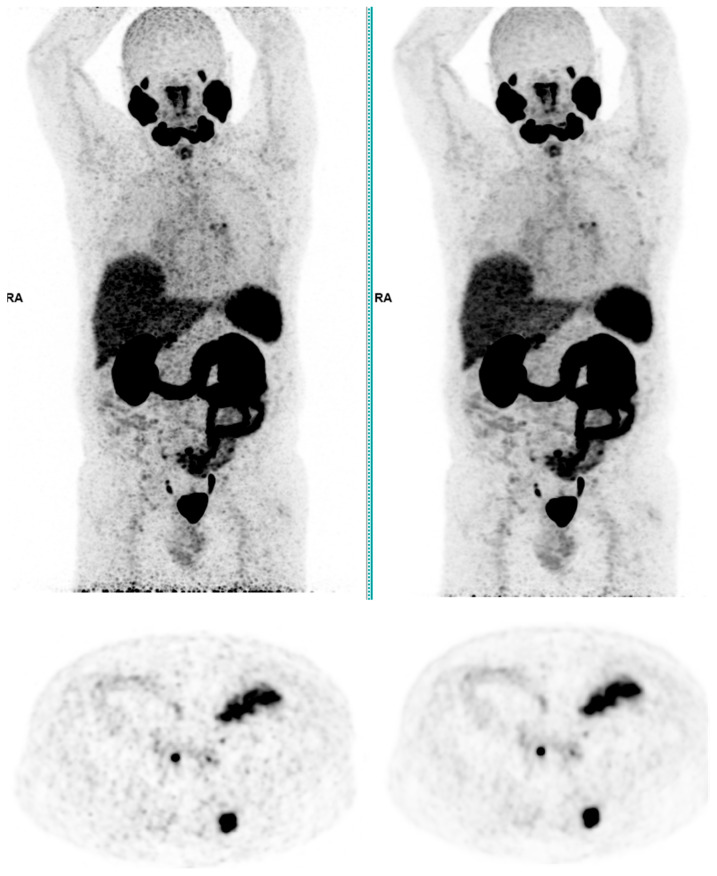
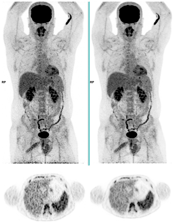
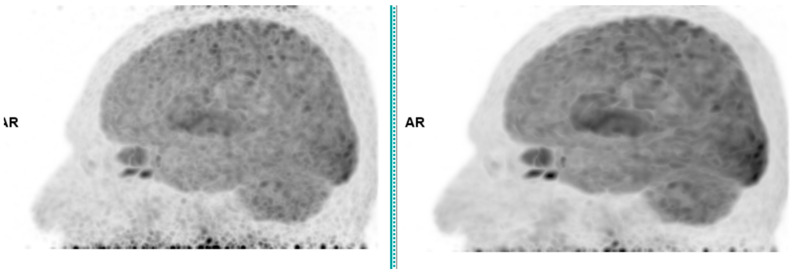
This study assesses the clinical deployment of SubtlePET™, a commercial AI-based denoising algorithm, across three radiotracers-18F-FDG, 68Ga-PSMA-11, and 18F-FDOPA-with the goal of improving image quality while reducing injected activity, technologist radiation exposure, and scan time. A retrospective analysis on a digital PET/CT system showed that SubtlePET™ enabled dose reductions exceeding 33% and time savings of over 25%. AI-enhanced images were rated interpretable in 100% of cases versus 65% for standard low-dose reconstructions. Notably, 85% of AI-enhanced scans received the maximum Likert quality score (5/5), indicating excellent diagnostic confidence and noise suppression, compared to only 50% with conventional reconstruction. The quantitative image quality improved significantly across all tracers, with SNR and CNR gains of 50-70%. Radiotracer dose reductions were particularly substantial in low-BMI patients (up to 41% for FDG), and the technologist exposure decreased for high-exposure roles. The daily patient throughput increased by an average of 4.84 cases. These findings support the robust integration of SubtlePET™ into routine clinical PET practice, offering improved efficiency, safety, and image quality without compromising lesion detectability.

 求助内容:
求助内容: 应助结果提醒方式:


